
Page 30 - MSAM-2-4
P. 30
Materials Science in Additive Manufacturing MAM for orthopedic bone plates: An overview
where mesenchymal cells transition into bone-forming they influence cellular behavior [6,13] . This interest stems
osteoblasts in a stable post-fracture environment . In partly from the extensive use of external implants in medical
[6]
contrast, secondary healing is a more common and efficient treatments. The choice of implant materials and design is
mechanism, encompassing a series of stages, namely, pivotal as they shape the mechanical microenvironment
hematoma formation, granulation tissue formation, callus at the fracture site [7,14] . Through tailored implants, one can
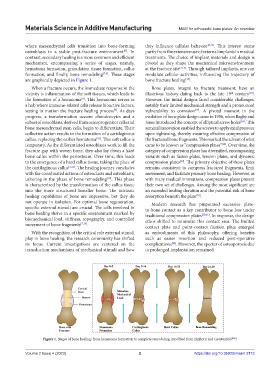
formation, and finally, bone remodeling [7,8] . These stages modulate cellular activities, influencing the trajectory of
are graphically depicted in Figure 1. bone fracture healing .
[15]
When a fracture occurs, the immediate response in the Bone plates, integral to fracture treatment, have an
[16]
th
vicinity is inflammation of the soft tissues, which leads to illustrious history dating back to the late 19 century .
the formation of a hematoma . This hematoma serves as However, the initial designs faced considerable challenges,
[8]
a hub where immune-related cells release bioactive factors, notably their limited mechanical strength and a pronounced
setting in motion the fracture healing process . As days vulnerability to corrosion . A pivotal moment in the
[9]
[17]
progress, a transformation occurs: chondrocytes and a evolution of bone plate design came in 1956, when Bagby and
subset of osteoblasts, derived from osteoprogenitor cells and Janes introduced the concept of elliptical screw holes . This
[18]
bone mesenchymal stem cells, begin to differentiate. Their seminal innovation enabled the screws to apply axial pressure
collective action results in the formation of a cartilaginous upon tightening, thereby ensuring effective compression of
callus, replacing the earlier hematoma . This soft callus is the fractured bone fragments. This marked the advent of what
[10]
temporary. As the differentiated osteoblasts work to fill the came to be known as “compression plates” . Over time, the
[18]
fracture gap with woven bone, they also lay down a hard category of compression plates has diversified, encompassing
bone callus within the periosteum. Over time, this leads variants such as fusion plates, tension plates, and dynamic
to the emergence of a hard callus tissue, taking the place of compression plates . The primary objective of these plates
[19]
the cartilaginous callus [3,11] . The healing journey concludes remains consistent: to compress fractured fragments, limit
with the coordinated actions of osteoclasts and osteoblasts, movement, and facilitate primary bone healing. However, as
ushering in the phase of bone remodeling . This phase with many medical innovations, compression plates present
[12]
is characterized by the transformation of the callus tissue their own set of challenges. Among the most significant are
into the more structured lamellar bone. The intrinsic an extended healing duration and the potential risk of bone
healing capabilities of bone are impressive, but they do resorption beneath the plate .
[20]
not operate in isolation. For optimal bone regeneration, Modern research has pinpointed excessive plate-
specific external stimuli are crucial. The cells involved in to-bone contact as a key contributor to bone loss under
bone healing thrive in a specific environment marked by traditional compression plates [20,21] . In response, the design
biomechanical load, stiffness, topography, and controlled ethos shifted to minimize this contact area. The limited
movement of bone fragments [7,13] . contact plate and point-contact fixation plate emerged
With the recognition of the critical role external stimuli as embodiments of this philosophy, offering benefits
play in bone healing, the research community has shifted such as easier insertion and reduced post-operative
its focus. Current investigations are centered on the complications . However, the specter of osteoporosis due
[22]
transduction mechanisms of mechanical stimuli and how to prolonged implantation remained.
Figure 1. Stages of bone healing: from hematoma formation to complete remodeling. Modified from Einhorn and Gerstenfeld [106] .
Volume 2 Issue 4 (2023) 2 https://doi.org/10.36922/msam.2113