
Page 88 - MSAM-3-2
P. 88
Materials Science in Additive Manufacturing Sustainable resin for coral restoration
a programmed ramp rate of 10°C/min. The sample hydrogel was employed to establish an ECM environment.
underwent a two-step cycle: This hydrogel, mimicking the collagen structure found in
(i) Heating from room temperature to 160°C and holding the ECM, has the capacity to create a 3D microenvironment
at 160°C for 1 min. that closely resembles native tissues. Among the various
30
(ii) Cooling from 160°C to −30°C and holding for another ultrashort self-assembling peptides tested, ultrashort self-
1 min. assembling peptide IIZK (Ac-Ile-Ile-Cha-Lys-NH ) was
2
Subsequently, the second cycle was used to determine selected. This peptide has previously been successfully
the Tg: utilized in the Laboratory for Nanomedicine to create
(i) Heating from −30°C to 160°C. diverse 3D in vitro cellular models. 31-33
(ii) Holding at 160°C for 1 min. The study utilized a 96-well assay plate (white plate,
(iii) Decreasing from 160°C to room temperature. clear bottom; Corning, USA). The vertical plate layout for
the experiment is structured as follows:
2.2.6. Cytotoxicity analysis
(i) The six columns on the left are designated for
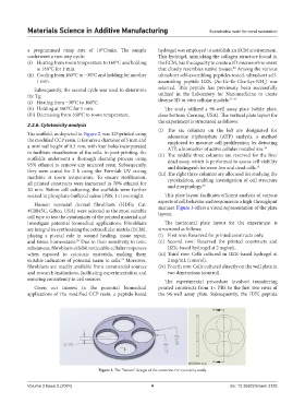
The scaffold, as depicted in Figure 2, was 3D-printed using adenosine triphosphate (ATP) analysis, a method
the modified CCP resin. It features a diameter of 5 mm and employed to measure cell proliferation by detecting
a minimal height of 0.3 mm, with four holes incorporated ATP, a biomarker of active cellular metabolism. 34
to facilitate visualization of the cells. In post-printing, the (ii) The middle three columns are reserved for the live/
scaffolds underwent a thorough cleaning process using dead assay, which is performed to assess cell viability
95% ethanol to remove any uncured resin. Subsequently, and distinguish between live and dead cells. 35
they were cured for 2 h using the Formlab UV curing (iii) The right three columns are allocated for studying the
machine at room temperature. To ensure sterilization, cytoskeleton, enabling investigation of cell structure
all printed constructs were immersed in 70% ethanol for and morphology. 36
30 min. Before cell culturing, the scaffolds were further
soaked in phosphate-buffered saline (PBS, 1×) overnight. This plate layout facilitates efficient analysis of various
aspects of cell behavior and responses in a high-throughput
Human neonatal dermal fibroblasts (HDFs; Cat.
#C0045C, Gibco, USA) were selected as the most suitable manner. Figure 3 offers a visual representation of the plate
layout.
cell type to test the cytotoxicity of the printed material and
investigate potential biomedical applications. Fibroblasts The horizontal plate layout for the experiment is
are integral in synthesizing the extracellular matrix (ECM), structured as follows:
playing a pivotal role in wound healing, tissue repair, (i) First row: Reserved for printed constructs only.
and tissue homeostasis. Due to their sensitivity to toxic (ii) Second row: Reserved for printed constructs and
28
substances, fibroblasts exhibit noticeable cellular responses IIZK-based hydrogel at 2 mg/mL.
when exposed to cytotoxic materials, making them (iii) Third row: Cells cultured in IIZK-based hydrogel at
suitable indicators of potential harm to cells. Moreover, 2 mg/mL (control).
29
fibroblasts are readily available from commercial sources (iv) Fourth row: Cells cultured directly on the well plate in
and research institutions, facilitating experimentation and two dimensions (control).
ensuring consistency in cell sources. The experimental procedure involved transferring
Given our interest in the potential biomedical printed constructs from 1× PBS to the first two rows of
applications of the modified CCP resin, a peptide-based the 96-well assay plate. Subsequently, the IIZK peptide
Figure 2. The “button” design of the construct for cytoxicity study.
Volume 3 Issue 2 (2024) 4 doi: 10.36922/msam.3125