
Page 92 - MSAM-3-2
P. 92
Materials Science in Additive Manufacturing Sustainable resin for coral restoration
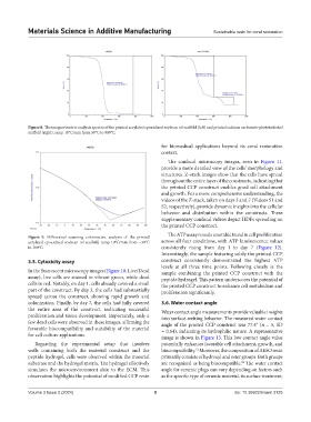
Figure 8. Thermogravimetric analysis spectra of the printed acrylated epoxidized soybean oil scaffold (left) and printed calcium carbonate-photoinitiated
scaffold (right), ramp 10°C/min from 50°C to 900°C.
for biomedical applications beyond its coral restoration
context.
The confocal microscopy images, seen in Figure 11,
provide a more detailed view of the cells’ morphology and
structures. Z-stack images show that the cells have spread
throughout the entire layer of the constructs, indicating that
the printed CCP construct enables good cell attachment
and growth. For a more comprehensive understanding, the
videos of the Z-stack, taken on days 3 and 7 (Videos S1 and
S2, respectively), provide dynamic insights into the cellular
behavior and distribution within the constructs. These
supplementary confocal videos depict HDFs spreading on
the printed CCP construct.
The ATP assay reveals a notable trend in cell proliferation
Figure 9. Differential scanning colorimetric analysis of the printed
acrylated epoxidized soybean oil scaffold, ramp 10°C/min from −30°C across all four conditions, with ATP luminescence values
to 100°C. consistently rising from day 1 to day 7 (Figure 12).
Interestingly, the sample featuring solely the printed CCP
3.5. Cytoxicity assay construct consistently demonstrated the highest ATP
levels at all three time points. Following closely is the
In the fluorescent microscopy images (Figure 10, Live/Dead sample combining the printed CCP construct with the
assay), live cells are stained in vibrant green, while dead peptide hydrogel. This pattern underscores the potential of
cells in red. Notably, on day 1, cells already covered a small the printed CCP construct to enhance cell metabolism and
part of the construct. By day 3, the cells had substantially proliferation significantly.
spread across the construct, showing rapid growth and
colonization. Finally, by day 7, the cells had fully covered 3.6. Water contact angle
the entire area of the construct, indicating successful Water contact angle measurements provide valuable insights
proliferation and tissue development. Importantly, only a into surface-wetting behavior. The measured water contact
few dead cells were observed in these images, affirming the angle of the printed CCP construct was 77.4° (n = 5, SD
favorable biocompatibility and suitability of the material = 0.64), indicating its hydrophilic nature. A representative
for cell culture applications. image is shown in Figure 13. This low contact angle value
Regarding the experimental setup that involves potentially enhances favorable cell attachment, growth, and
wells containing both the material construct and the biocompatibility. Moreover, the composition of AESO resin
51
peptide hydrogel, cells were observed within the material primarily consists of hydroxyl and ester groups. Both groups
substrate and the hydrogel matrix. The hydrogel effectively are recognized as being biocompatible. The water contact
52
simulates the microenvironment akin to the ECM. This angle for ceramic plugs can vary depending on factors such
observation highlights the potential of modified CCP resin as the specific type of ceramic material, its surface treatment,
Volume 3 Issue 2 (2024) 8 doi: 10.36922/msam.3125