
Page 93 - MSAM-3-2
P. 93
Materials Science in Additive Manufacturing Sustainable resin for coral restoration
Figure 10. Fluorescent microscopy images of human dermal fibroblasts spreading on the printed calcium carbonate-photoinitiated construct on days 1,
3, and 7, from left to right. Scale bars: 150 µm.
Figure 11. Confocal images of human dermal fibroblasts spreading on the printed calcium carbonate-photoinitiated construct on days 1, 3, and 7, from
left to right. Scale bars: 100 µm.
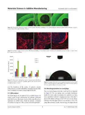
1200000
DAY 1
1000000
DAY 3
ATP Luminescence (RLU) 600000
DAY 7
800000
400000
200000
0
Construct Construct+Peptide Peptide 2D
Figure 12. Adenosine triphosphate levels of human dermal fibroblasts
cultured in construct, construct + peptide, peptide, and two dimensions Figure 13. Image of the water contact angle measurement for the printed
measured at days 1, 3, and 7.
calcium carbonate-photoinitiated scaffold. The dimension of the needle is
0.51 mm, and the volume of the water is 2.2 µL.
and the cleanliness of the surface. In general, ceramic
surfaces tend to be also hydrophilic, meaning that they often 3.8. Microfragmentation on coral plugs
have a relatively low water contact angle below 90°.
The coral microfragments at day 1 and day 20 are depicted
3.7. SEM analysis in Figure 15. The coral plugs were randomly distributed
The SEM images of the printed CCP scaffold (Figure 14) inside the water tank to minimize the variables, and they
reveal a well-mixed combination of calcium carbonate were assembled solely for imaging purposes. After day 7,
and AESO resin. The larger chunks with a smooth surface marine life started settling on the CCP and ceramic plugs.
represent the AESO resin, while the smaller crystalline Subsequently, the algae started to accumulate on the coral
structures correspond to the calcium carbonate particles. plugs after around 2 weeks. Interestingly, the algae almost
Volume 3 Issue 2 (2024) 9 doi: 10.36922/msam.3125