
Page 21 - OR-1-1
P. 21
A B C
D E F
G H
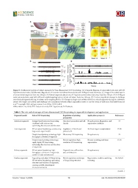
Figure 8. Development analysis of retinal organoids by three-dimensional (3D) bioprinting. (A) Schematic diagram of a microelectrode array with 3D
liquid metal electrodes. (B) Schematic diagram of a 3D anterior retinal membrane device with 3D liquid metal electrodes. (C) Image of the retinal layer in
a human retinal organoid. Scale bar, 200 μm. (D) Retinal organoids placed on a 3D liquid metal microelectrode array. Scale bar, 500 μm. (E) A 3D liquid
metal microelectrode array with different heights targeting various retinal cell layers. Scale bar, 300 μm. (F) Contour map shows changes in discharge
rate of retinal organoids during 16 culture cycles targeting RGCs. (G) Changes in single-unit activity of RGCs in retinal organoids during the cultivation
period. (H) Single-unit activity and discharge rate comparison between retinal organoids at week 11 and the retina of wild mice. Reprinted from Lee
et al. Copyright 2024, with permission from Wiley-VCH GmbH.
87
Abbreviations: Au: Gold; ITO: Indium tin oxide; LM: Liquid metal; Pt: Platinum.
Table 1. The role and advantages of three-dimensional (3D) bioprinting in organoid development and application
Organoid model Role of 3D bioprinting Regulation of printing Application prospects References
parameters
Intestinal organoids Syringe-based extrusion bioprinting Structural parameters and cell Drug discovery, diagnostics, and 33
combined with microscopy; density regenerative medicine
optimizing the structure and function
Liver organoids 3D extrusion bioprinting; constructing Regulation of bioink and Artificial organ transplantation 37,38
large-scale organ models printed cells
Droplet-based printing technology; high- Microarray 3D bioprinting Drug discovery 39
throughput cultivation of organoids
Droplet-based printing technology; Bioink regulation; high- Disease modeling and tissue 57,78
rapid, digital 3D bioprinting; resolution 3D bioprinting regeneration
optimizing the structure and function
of organoids
Kidney organoids 3D extrusion bioprinting; high- Organoid size, cell number, Drug discovery 40
throughput cultivation of organoids; and conformation
control of scale and structure
Cardiac organoids Expanding embedded 3D bioprinting; Bioink regulation and design Regenerative medicine and drug 43,44,59,61
3D extrusion bioprinting; optimizing of the printing structure screening
structure and function of organoids,
creating suitable physiological
microenvironments
(Cont’d...)
Volume 1 Issue 1 (2025) 13 doi: 10.36922/OR025040004