
Page 17 - OR-1-1
P. 17
A
B C
D E F G H I
J K
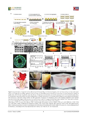
Figure 5. Vascularization of organoids by three-dimensional (3D) bioprinting. (A) Schematic diagram of the “sacrificial writing into functional tissue”
process. (B) Design plan for cardiac organoids. Scale bar, 50 μm. (C) Branching vascular networks by 3D printing. (D) Cross-sectional cell viability of
cardiac tissue after 24 hours of perfusion. Scale bar, 500 μm. (E) Spontaneous contraction and electrical pacing (1 and 2 Hz) of printed cardiac tissue.
(F) Spontaneous contractions before and after administration of 10 μM isoproterenol. (G) Average systolic frequency under isoproterenol treatment.
(H) Spontaneous contraction patterns before and after application of heptanol. (I) Maximum peak-to-peak shrinkage amplitude under heptanol
treatment. (J) Schematic diagram of the structure of an artificial heart model. (K) Images of printed heart tissue. Scale bar, 5 mm. Reprinted from Skylar-
61
Scott et al. Copyright 2019, with permission from the American Association for the Advancement of Science.
Abbreviations: CAD: Coronary artery disease; CDM: Cardiomyocyte differentiation medium; CDMC: CDM and 5 μM CHIR99021; CDMI: CDM
and 2 μM iWR1; CMM: Cardiomyocyte maturation medium; Ctrl: Control; Diag: Diagonal; EBs: Embryoid bodies; ECM: Extracellular matrix;
EthD-1: Ethidium homodimer-1; Isoprot: isoproterenol; LAD: Left anterior descending artery; mTeSR: Refers to the name of a culture medium produced
by STEMCELL Technologies; PVA: Polyvinyl Alcohol; Sept: Septum; vent: Ventricle; 2D: Two-dimensional.
Volume 1 Issue 1 (2025) 9 doi: 10.36922/OR025040004