
Page 15 - OR-1-1
P. 15
A B
C D
E
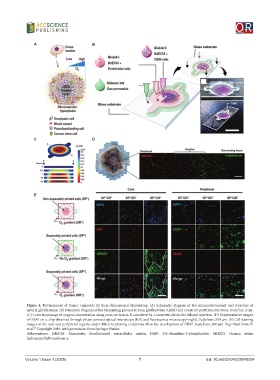
Figure 4. Development of tumor organoids by three-dimensional bioprinting. (A) Schematic diagram of the microenvironment and structure of
natural glioblastoma. (B) Schematic diagram of the bioprinting process to form glioblastoma (GBM) and construct partition structures. Scale bar, 2 cm.
(C) Color map image of oxygen concentration along cross-section A-A’, simulated by a computer, shows the delayed injection. (D) Representative images
of GBM-on-a-chip observed through phase contrast optical microscopy (left) and fluorescence microscopy (right). Scale bars, 500 μm. (E) Cell staining
images of the core and peripheral regions under different printing conditions show the development of GBM. Scale bars, 200 μm. Reprinted from Yi
54
et al. Copyright 2019, with permission from Springer Nature.
Abbreviations: BdECM: Biomimetic decellularized extracellular matrix; DAPI: 4’,6-diamidino-2-phenylindole; SHMT2: Human serine
hydroxymethyltransferase 2.
Volume 1 Issue 1 (2025) 7 doi: 10.36922/OR025040004