
Page 12 - OR-1-1
P. 12
A B
C
D E
F
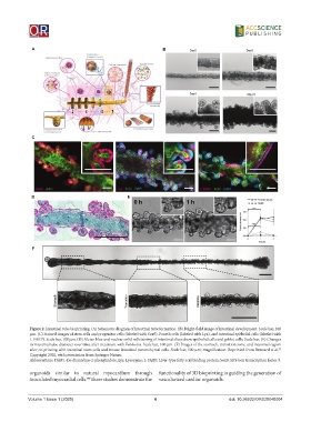
Figure 2. Intestinal tube bioprinting. (A) Schematic diagram of intestinal tube formation. (B) Bright-field image of intestinal development. Scale bar, 200
μm. (C) Stained images of stem cells and progenitor cells (labeled with Sox9), Paneth cells (labeled with Lyz), and intestinal epithelial cells (labeled with
L-FABP). Scale bar, 100 μm. (D) Alcian blue and nuclear solid red staining of intestinal slices show epithelial cells and goblet cells. Scale bar. (E) Changes
in intestinal tube diameter over time after treatment with Forskolin. Scale bar, 100 μm. (F) Images of the stomach, transition zone, and intestinal region
33
after co-printing with intestinal stem cells and mouse intestinal mesenchymal cells. Scale bar, 500 μm; magnification. Reprinted from Brassard et al.
Copyright 2020, with permission from Springer Nature.
Abbreviations: DAPI: 4’,6-diamidino-2-phenylindole; Lyz: Lysozyme; L-FABP: Liver-type fatty acid binding protein; Sox9: SRY-box transcription factor 9.
organoids similar to natural myocardium through functionality of 3D bioprinting in guiding the generation of
inoculated myocardial cells. These studies demonstrate the vascularized cardiac organoids.
44
Volume 1 Issue 1 (2025) 4 doi: 10.36922/OR025040004