
Page 13 - OR-1-1
P. 13
3.2. Organoids from the musculoskeletal system networks, and decellularised gastric extracellular matrix,
all constructed via bioprinting. The highly vascularised
53
Bone tissue is a crucial organ in the human body, approach achieved through 3D bioprinting provides a
responsible for various important functions. Due to
45
its inherent structural complexity and diverse cellular superior simulation of the tumor microenvironment. In
composition, developing bone organoids is extremely addition, the decellularised gastric extracellular matrix
challenging. However, 3D printing technology can provides a suitable microenvironment for gastric cancer
46
reconstruct these complex structures. 47,48 For example, organoids, facilitating the study of tumor-extracellular
matrix interactions. Similarly, using 3D bioprinting,
Zhang et al. developed a Haversian-simulated bone patient-derived glioblastoma cells, vascular endothelial
49
structure scaffold, which includes cortical and cancellous cells, and brain tissue decellularized extracellular matrix
bone structures, as well as Haversian and Volkmann tubes. were printed to form a tumor-matrix concentric ring
3D bioprinting integrates cells into specific bone tissue structure. These tumor organoids maintain a radial
54
structures, providing a clear direction for the development oxygen gradient, reflecting the structural properties of
of bone organoids. A team led by Prof Jiacan Su at Shanghai natural tumors (Figure 4). Importantly, the development
University mixed inorganic hydroxyapatite with organic of these specific malignant tumor organoids offers
polymers to create a bioink, which was printed using great potential for patient treatment. High-throughput
digital light processing (DLP)-based 3D bioprinting to culture of tumor organoids is essential for evaluating and
create a bone organoid scaffold. This study showed that developing anti-tumor drugs. At present, Matrigel is the
50
the resulting construct provides functional mechanical primary matrix material used for tumor organoid culture;
support, supports long-term cell culture, and exhibits self- however, its limited mechanical properties and variability
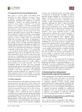
mineralization ability both in vitro and in vivo. After in vivo in tumor models present challenges. 3D bioprinting offers
cultivation, the organoids further developed and displayed a more suitable microenvironment, enabling repeatable
a trabecular bone structure (Figure 3). This study offers and high-throughput cultivation of tumor organoids.
a clear strategy for developing bone organoids using 3D Research has shown that colorectal cancer organoids can
bioprinting, influencing the development of bone tissue- grow successfully in composite bioinks, allowing for the
related biomedical research. In another study, the hydrogel generation of large numbers of organoid microspheres for
microspheres encapsulating bone marrow mesenchymal anti-tumor drug screening. 55
stem cells were prepared in batches through the DLP-based
bioprinting technology. These microspheres were cultured In this section, we have discussed the development
in the cartilage-forming medium for 3 weeks, leading to of various organoids by 3D bioprinting. The role of 3D
the development of osteo-callus organoids. The in vitro bioprinting technology in organoid development is also
51
development pattern of these osteo-callus organoids highlighted. The accuracy and flexibility of 3D bioprinting
resembles natural endochondral ossification. When used to are crucial in enhancing organoid structures, creating ideal
repair large bone defects in vivo, the osteo-callus organoids microenvironments for organoid growth, high-throughput
accelerate in situ bone regeneration. 3D bioprinting cultivation, and enabling the scaling-up of organoids.
provides a desirable strategy for the preparation of bone- 4. Harnessing three-dimensional
related organoids, which not only simulate the structure
and developmental patterns of bone tissue but also facilitate bioprinting for organoid development
the large-scale production of bone-related organoids. Various organoids have been integrated with 3D bioprinting
to develop different tissues and organs. The precision
3.3. Tumor organoids
and flexibility of 3D bioprinting play an important role
Tumors have long been a major medical challenge, seriously in improving organoid structures, creating optimal
threatening human life and health. Recent research has microenvironments for organoid growth, facilitating high-
shown that tumor organoids derived from patients retain a throughput cultivation, and enabling organoid enlargement.
high fidelity to the original tumors, providing an excellent In this section, we review the advantages of 3D bioprinting
model for personalized treatment and the study of tumor in organoid development, including the optimization
pathology. However, limitations in tumor organoid of organoid structure and function, the vascularization
52
development persist, including the lack of important of organoids, and strategies for enhancing organoid
extracellular matrix components, the challenge of simulating development and application through 3D bioprinting.
unique physiological microenvironments, and the need for
high-throughput culture systems. The application of 3D 4.1. Optimization of organoid structure and function
bioprinting aims to address these issues. For example, one via three-dimensional bioprinting
study developed a vascularized organoid model composed One of the key features of organoids is their ability to
of patient-derived gastric cancer organoids, endothelial simulate the structures and functions of real tissues. These
56
Volume 1 Issue 1 (2025) 5 doi: 10.36922/OR025040004