
Page 20 - OR-1-1
P. 20
A B
C
D E
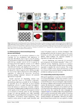
Figure 7. Development of neural organoids by three-dimensional bioprinting. (A) Schematic diagram of magnetic enhancement of neural organoids and
bright-field image of neural organoids after magnetic enhancement. Scale bar, 1,000 μm. (B) Schematic diagram of the spatially patterned organ transfer
platform. (C) Fusion and free assembly of organoids. (D) Fusion of neuromorphic organoid and diffuse pontine glioma organoid. (E) Bright-field images
and immunofluorescence staining of organoid complexes. Reprinted from Roth J. G. et al. Copyright 2021, with permission from Springer Nature.
71
Abbreviation: EGFP: Enhanced green fluorescent protein.
5.3. Enhancing three-dimensional bioprinting control 3D printed constructs, has been developed. This
84
devices and methods printing method allows for the precise control of structure
The optimization of 3D bioprinting equipment is also formation, as well as the accurate positioning of printed
important for the development and application of spheres, which facilitates the controlled assembly of
organoids. Microfluidic chips designed using 3D printing organoids into large-scale organ models.
can explore interactions between organoids and endothelial Acoustic bioprinting can manipulate the generation
cells forming a vascular network, which is essential for and arrangement of individual organoids. Chen et al.
85
86
developing functional organoids. Salmon et al. utilized employed acoustic bioprinting equipment to construct
82
this platform to promote interactions between brain 3D structures with precisely arranged tumors and healthy
organoids and vascular cells, leading to the formation of organoids to simulate the colon of patients. This model
neurovascular organoids. The 3D bioprinting platform can was used to analyze tumor spread and invasion, as well
be adapted for various organoid types, offering strategies as to conduct drug screening, thereby assisting healthcare
for vascularized organoid development and enabling the providers in making treatment decisions.
exploration of organoid-cell interactions. Specifically,
adjusting printing equipment enhances the potential 5.4. Incorporating monitoring measures
applications of organoids. Although the application of organoids in the biomedical field
Several improved 3D bioprinting systems have is becoming increasingly common, there remains a lack of
shown promising results in microsphere preparation, monitoring methods for certain physiological characteristics
demonstrating significant potential in the development of their development. One study designed a high-resolution
83
of organoids. Xie et al. developed an electro-assisted 3D printing combined with liquid metal microelectrodes to
bioprinting method to prepare hydrogel microspheres. monitor the development of ganglion cells in early to mid-
87
Under the influence of electrostatic attraction, uniform- stage retinal organoids. This device can accurately locate the
sized photo-crosslinked gelatin methacryloyl microspheres inner layer of the retina within the organoid and record signals
can be rapidly printed. In addition, the separation method within the retina, thus avoiding the risk of damaging the
of microspheres is relatively gentle on cells, minimizing organoid (Figure 8). In addition, inkjet 3D printing has been
damage and reducing printing costs while improving used to prepare microelectrode arrays capable of recording
printing efficiency, providing valuable strategies for organoid electrophysiological signals from cortical organoids, again
development. Furthermore, hybrid bioprinting, which avoiding damage to the organoids. Moreover, 3D-printed
88
combines suction power to apply liquid absorption and electrochemical porous plates have been applied to monitor
Volume 1 Issue 1 (2025) 12 doi: 10.36922/OR025040004