
Page 101 - OR-1-2
P. 101
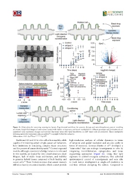
Figure 11. Mini-colons for screening tumorigenic factors. Experimental workflows for systemic therapy and microbiota/dietary pattern modeling
            are shown. Bright-field images of mini-colons treated with vehicle or tiopronin, and tumor multiplicity in different genotypes and pretreatments are
            quantified. Gene expression changes and enriched functional terms after Gpx2 knockdown in AKP tumor cells are presented. Tumor multiplicity
                                            85
            following metabolite treatments is also quantified.  Copyright © 2024 The author(s).
                            fl/fl
                                        fl/fl
            Abbreviation: AKP: Apc Kras LSL-G12D/+ Trp53 .
               Traditional 2D and 3D in vitro cell culture models, while   high-resolution analyses of cellular dynamics in terms
            capable of mimicking certain simple cancer cell behaviors,   of temporal and spatial resolution and are also costly  in
            have limitations in  simulating complex tissue structures   terms of resources. Lorenzo-Martín  et al.  developed a
                                                                                                 85
            and the process of cancer development.  Current organoid   “mini-colon” that can undergo tumorigenesis  in vitro by
                                            83
            models, although considered a bridge between in vitro and   integrating microfabrication, optogenetics, and tissue
            in vivo models, are limited by their closed structure, short   engineering  approaches  (Figure  11).  Using blue  light
            lifespan, lack of stable tissue architecture, and inability   exposure to control oncogene activation, they achieved
            to generate hybrid tissues composed of both healthy and   spatiotemporal control of tumorigenesis and were able
                      84
            cancer cells.  These limitations mean that cancer research   to track tumor development at single-cell resolution in
            still relies heavily on animal models, which cannot provide   real-time without disrupting the culture. Compared to


            Volume 1 Issue 2 (2025)                         16                           doi: 10.36922/OR025040005