
Page 97 - OR-1-2
P. 97
A C
D
B
E
F G
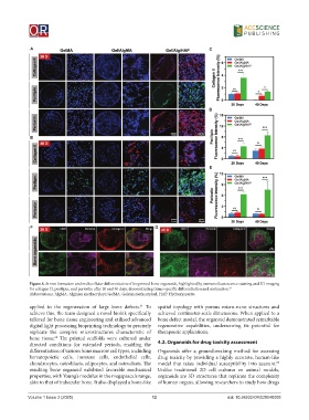
Figure 8. In vivo formation and multicellular differentiation of bioprinted bone organoids, highlighted by immunofluorescence staining and 3D imaging
for collagen II, perilipin, and periostin after 20 and 40 days, demonstrating tissue-specific differentiation and maturation. 61
Abbreviations: AlgMA: Alginate methacryloyl; GelMA: Gelatin methacryloyl; HAP: Hydroxyapatite.
applied to the regeneration of large bone defects. To spatial topology with porous micro-nano structures and
61
achieve this, the team designed a novel bioink specifically achieved centimeter-scale dimensions. When applied to a
tailored for bone tissue engineering and utilized advanced bone defect model, the organoid demonstrated remarkable
digital light processing bioprinting technology to precisely regenerative capabilities, underscoring its potential for
replicate the complex microstructures characteristic of therapeutic applications.
bone tissue. The printed scaffolds were cultured under
62
directed conditions for extended periods, enabling the 4.2. Organoids for drug toxicity assessment
differentiation of various bone marrow cell types, including Organoids offer a groundbreaking method for assessing
hematopoietic cells, immune cells, endothelial cells, drug toxicity by providing a highly accurate, human-like
chondrocytes, osteoblasts, adipocytes, and osteoclasts. The model that takes individual susceptibility into account.
63
resulting bone organoid exhibited favorable mechanical Unlike traditional 2D cell cultures or animal models,
properties, with Young’s modulus in the megapascals range, organoids are 3D structures that replicate the complexity
akin to that of trabecular bone. It also displayed a bone-like of human organs, allowing researchers to study how drugs
Volume 1 Issue 2 (2025) 12 doi: 10.36922/OR025040005