
Page 96 - OR-1-2
P. 96
A B D
C
E F H
I
G
J
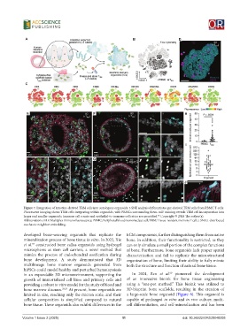
Figure 7. Integration of intestine-derived TRM cells into autologous organoids. t-SNE analysis differentiates gut-derived TRM cells from PBMC T-cells.
Fluorescent imaging shows TRM cells integrating within organoids, with PBMCs surrounding them. mIF staining reveals TRM cell incorporation into
larger and smaller organoids. Immune cell counts and epithelial-to-immune cell ratios are quantified. Copyright © 2024 The author(s).
46
Abbreviations: mIF: Multiplex immunofluorescence; PBMC: Peripheral blood mononuclear cell; TRM: Tissue-resident memory T cell; t-SNE: t-distributed
stochastic neighbor embedding.
developed bone-weaving organoids that replicate the ECM components, further distinguishing them from native
mineralization process of bone tissue in vitro. In 2022, Xie bone. In addition, their functionality is restricted, as they
et al. constructed bone callus organoids using hydrogel can only simulate a small portion of the complex functions
57
microspheres as stem cell carriers, a novel method that of bone. Furthermore, bone organoids lack proper spatial
mimics the process of endochondral ossification during characterization and fail to replicate the microstructural
bone development. A study demonstrated that 3D organization of bone, limiting their ability to fully mimic
multilineage bone marrow organoids generated from both the structure and function of natural bone tissue.
hiPSCs could model healthy and perturbed hematopoiesis
60
in an expandable 3D microenvironment, supporting the In 2024, Ren et al. pioneered the development
growth of immortalized cell lines and primary cells and of an innovative bioink for bone tissue engineering
providing a robust in vitro model for the study of blood and using a “one-pot method.” This bioink was utilized to
bone marrow diseases. 58,59 At present, bone organoids are 3D-bioprint bone scaffolds, resulting in the creation of
limited in size, reaching only the micron scale, and their a large-scale bone organoid (Figure 8). This organoid is
cellular composition is simplified compared to natural capable of prolonged in vitro and in vivo culture, multi-
bone tissue. These organoids also exhibit differences in the cell differentiation, and self-mineralization and has been
Volume 1 Issue 2 (2025) 11 doi: 10.36922/OR025040005