
Page 42 - TD-1-1
P. 42
Tumor Discovery A bioinformatics analysis of PD-1 in cancers
A B
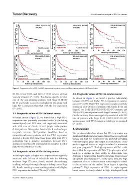
Figure 1. Prognostic value of PD-1 mRNA expression in gastric cancer and liver cancer patients. HR, hazard ratio.
III-IV), Grade II/III, and AJCC-T II/III cancers without 3.5. Prognostic values of PD-1 in ovarian cancer
vascular invasion (P < 0.05). The disease-specific survival As shown in Figure 4, we found a positive relationship
rate of the non-drinking patients with Stage II-III/III/ between OS/PFS and higher PD-1 expression in ovarian
III-IV and Grade I cancers was higher in the group with cancer (P < 0.05). High PD-1 expression was also positively
high PD-1 expression than that with the low expression correlated with OS rate of patients with Grade II+III and
(P < 0.05). Stage II (or II+III/III/IV/III+IV/II+III+IV) cancers, and
3.3. Prognostic values of PD-1 in breast cancer PFS (or PPS) rate of patients with Stage II cancer (P < 0.05).
On the contrary, there was negatively associated with PFS
In breast cancer (Figure 2), we found that a high PD-1 rate of patients with Stage II+III (III/III+IV/II+III+IV)
expression was positively associated with OS (including serous cancer with TP53 mutation (wild-type or mutated)
mesenchymal) and RFS rates, and negatively associated (P < 0.05).
with PPS rate of Grade II and lymph node-positive
(LN+) patients. ER-negative, luminal (A, B, and androgen 4. Discussion
receptor), intrinsic Her2-positive, basal-like, basal or The previous studies have shown that PD-1 expression was
mesenchymal cancer patients with low PD-1 expression significantly higher in breast cancer tissues than normal breast
showed a shorter RFS time than those with high PD-1 tissues, and high PD-1 expression was positively correlated
expression, but a negative association between PD-1 with histological grade and lymph node metastasis. These
expression and the RFS of progesterone receptor-positive results suggested that PD-1 might be related to metastasis
patients was present (P < 0.05). and poor prognosis . The high expression of PD-1 could
[12]
also inhibit the expression of CD8+ T lymphocytes, which
3.4. Prognostic values of PD-1 in lung cancer
leads to a decline in their ability to differentiate into
According to Figure 3, a higher PD-1 expression was negatively cytotoxic T lymphocytes, thereby promoting breast cancer
associated with OS rate of individuals with the following cell growth and metastasis . At the same time, the high
[18]
attributes: Stage I/II cancer, female, received chemotherapy, expression of PD-1 in breast cancer tissues might be related
smoking, and negative surgical margins in lung cancer. Stage to the activation of the nuclear factor kappa B signaling
N1, female, smoking or non-smoking patients with higher pathway . High expression of PD-1 in triple-negative
[19]
PD-1 expression had a short PPS rate (P < 0.05). breast cancer is closely related to high histological levels
Volume 1 Issue 1 (2022) 3 https://doi.org/10.36922/td.v1i1.59