
Page 20 - TD-3-1
P. 20
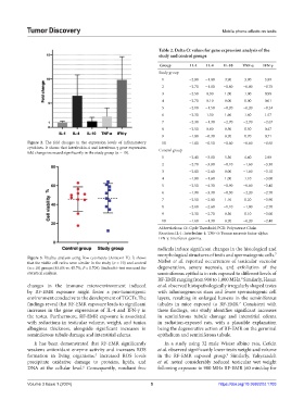
Tumor Discovery                                                             Mobile phone effects on testis


                                                               Table 2. Delta Ct values for gene expression analysis of the
                                                               study and control groups
                                                                Group       IL‑1  IL‑4   IL‑10  TNF‑α   IFN‑γ
                                                               Study group
                                                                1          −2.80  −1.00  3.90    3.90    3.89
                                                                2          −2.70  −1.80  −0.80  −0.80   −0.78
                                                                3          −2.50  0.90   1.00    1.00    0.98
                                                                4          −2.70  0.10   0.00    0.00    0.01
                                                                5          −2.10  −1.50  −0.20  −0.20   −0.24
                                                                6          −2.30  1.50   1.60    1.60    1.57
                                                                7          −2.30  −1.30  −2.70  −2.70   −2.67
                                                                8          −2.50  0.00   0.50    0.50    0.47
                                                                9          −1.80  −0.30  0.70    0.70    0.71
            Figure  2.  The  fold  changes  in the  expression  levels  of inflammatory   10  −1.80  −0.50  −0.60  −0.60  −0.65
            cytokines. It shows that interleukin-4 and interferon-γ gene expression   Control group
            fold change increased significantly in the study group (n = 10).
                                                                1          −2.40  −3.80  5.30    4.40    2.90
                                                                2          −2.70  −3.80  −0.10  −1.60   −3.30
                                                                3          −2.80  −2.60  0.00   −1.60   −3.10
                                                                4          −1.80  −1.60  1.00    1.10   −3.00
                                                                5          −2.50  −4.30  −0.90  −0.60   −3.40
                                                                6          −1.90  −3.30  −0.30  −2.20   −2.70
                                                                7          −2.50  −2.00  1.10    0.20   −3.90
                                                                8          −2.60  −2.60  −1.10  −1.00   −2.70
                                                                9          −2.30  −2.70  0.30    0.10   −3.00
                                                                10         −1.60  −1.30  0.70   −0.20   −2.40
                                                               Abbreviations: Ct: Cycle Threshold; PCR: Polymerase Chain
                                                               Reaction; IL-1: Interleukin-1; TNF-α: Tumor necrosis factor-alpha;
                                                               IFN-γ: Interferon-gamma.
                                                               radicals induce significant changes in the histological and
                                                               morphological structures of testis and spermatogenic cells.
                                                                                                             7
            Figure 3. Vitality analysis using flow cytometry (Annexin V). It shows
            that the viable cell ratios were similar in the study (n = 10) and control   Nisbet  et al. reported  occurrences of  testicular vacuolar
            (n = 10) groups (43.6% vs. 45.7%, P = 0.709). Student’s t-test was used for   degeneration, severe necrosis, and exfoliation of the
            statistical analysis.                              seminiferous epithelia in rats exposed to different levels of
                                                               RF-EMR ranging from 900 to 1,800 MHz.  Similarly, Hasan
                                                                                                8
            changes in the immune microenvironment induced     et al. observed histopathologically irregularly shaped testes
            by  RF-EMR exposure  might  foster  a pro-tumorigenic   with inhomogeneous sizes and fewer spermatogenic cell
            environment conducive to the development of TGCTs. The   layers, resulting in enlarged lumens in the seminiferous
            findings reveal that RF-EMR exposure leads to significant   tubules in mice exposed to RF-EMR.  Consistent with
                                                                                               7
            increases in the gene expressions of IL-4 and IFN-γ in   these findings, our study identifies significant increases
            the testes. Furthermore, RF-EMR exposure is associated   in seminiferous tubule damage and interstitial edema
            with reductions in testicular volume, weight, and tunica   in radiation-exposed rats, with a plausible explanation
            albuginea  thickness,  alongside  significant  increases  in   being the degenerative action of RF-EMR on the germinal
            seminiferous tubule damage and interstitial edema.  epithelium and seminiferous tubule.
              It has been demonstrated that RF-EMR significantly   In a study using 32  male Wistar albino rats, Cetkin
            weakens antioxidant enzyme activity and increases ROS   et al. observed significantly lower testis weight and volume
            formation in living organisms.  Increased ROS levels   in the RF-EMR exposed group.  Similarly, Yahyazadeh
                                      5
                                                                                          9
            precipitate oxidative damage to proteins, lipids, and   et  al.  noted  considerably  reduced  testicular  wet  weight
            DNA at the cellular level.  Consequently, resultant free   following exposure to 900 MHz RF-EMR (60 min/day for
                                  6
            Volume 3 Issue 1 (2024)                         5                          https://doi.org/10.36922/td.1703