
Page 22 - TD-3-1
P. 22
Tumor Discovery Mobile phone effects on testis
specific cytokines, including IL-4, IFN-γ, growth factors,
and oncogenes. Other studies have suggested that IL-4-
17
mediated signaling through STAT contributes to tumor
development within the tumor microenvironment. 18-21
Indeed, studies have specifically indicated that dysregulated
IL-4 formation is associated with various cancer types. 21,22
IL-4 is a versatile cytokine and vital for regulating
the immune system. Upon IL-4 binding to its cytokine
22
receptor, the ensuing activation of cell growth mediators,
resistance to apoptosis, gene activation, and differentiation
occur. The significance of IL-4 as a promoter of tumor-
22
initiating/cancer stem cell (CSC)-like cells has been
demonstrated across various cancers. Elevated levels of IL-4
(generally generated by tumor-infiltrating lymphocytes)
have been verified in advanced-stage prostate cancer
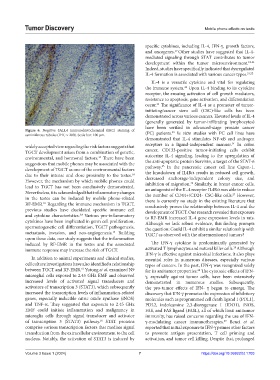
Figure 6. Negative SALL4 immunohistochemical (IHC) staining of 23
seminiferous tubules (IHC × 200). Scale bar: 100 μm. (PC) patients. In vitro studies with PC cell lines have
demonstrated that IL-4 stimulates NF-κB and androgen
24
widely accepted view regarding the risk factors suggests that receptors in a ligand-independent manner. In colon
TGCT development arises from a combination of genetic, cancer, CD133-positive tumor-initiating cells exhibit
environmental, and hormonal factors. There have been autocrine IL-4 signaling, leading to the upregulation of
13
suggestions that mobile phones may be associated with the the anti-apoptotic protein Survivin, a target of the STAT-6
25
development of TGCT as one of the environmental factors pathway. In the pancreatic cancer cell line Capan-1,
the knockdown of IL4Rα results in reduced cell growth,
due to their intense and close proximity to the testes. decreased anchorage-independent colony size, and
14
However, the mechanism by which mobile phones could inhibition of migration. Similarly, in breast cancer cells,
26
lead to TGCT has not been conclusively demonstrated. an antagonist of the IL-4 receptor IL4Rα was able to reduce
Nevertheless, it is acknowledged that inflammatory changes the number of CD44+/CD24- CSC-like cells. However,
27
in the testes can be induced by mobile phone-related there is currently no study in the existing literature that
RF-EMR. Regarding the immune mechanism in TGCT, conclusively proves the relationship between IL-4 and the
10
previous studies have elucidated specific immune cell development of TGCT. Our research revealed that exposure
and cytokine characteristics. Various pro-inflammatory to RF-EMR increased IL-4 gene expression levels in rats.
3,4
cytokines have been implicated in germ cell proliferation, Although we lack robust evidence, this finding prompts
spermatogenetic cell differentiation, TGCT pathogenesis, the question: Could IL-4 exhibit a similar relationship with
metastasis, invasion, and neo-angiogenesis. Building TGCT as observed with the aforementioned tumors?
15
upon these data, our study suggests that the inflammation
induced by RF-EMR in the testes and the associated The IFN-γ cytokine is predominantly generated by
28
immune response may increase the risk of TGCT. activated T lymphocytes and natural killer cells. Although
IFN-γ is effective against microbial infections, it also plays
In addition to animal experiments and clinical studies, essential roles in numerous diseases, especially various
cell culture investigations have also identified a relationship types of cancers. In the past, IFN-γ was recognized solely
between TGCT and RF-EMR. Yutong et al. examined N9 for its antitumor properties. The cytotoxic effects of IFN-
16
28
microglial cells exposed to 2.45 GHz EMF and observed γ, especially against tumor cells, have been extensively
increased levels of activated signal transducers and demonstrated in numerous studies. Subsequently,
activators of transcription 3 (STAT3), which subsequently the pro-tumor effects of IFN- γ began to emerge. The
increased the transcription levels of inflammation-related discovery that IFN-γ promotes the expression of inhibitory
genes, especially inducible nitric oxide synthase (iNOS) molecules such as programmed cell death ligand 1 (PDL1),
and TNF-α. They suggested that exposure to 2.45 GHz PDL2, indoleamine 2,3-dioxygenase 1 (IDO1), iNOS,
EMF could initiate inflammation and malignancy in FAS, and FAS ligand (FASL), all of which limit antitumor
microglia cells through signal transducer and activator immunity, has raised concerns regarding the use of IFN-
16
of transcription 3 (STAT3) pathway. STAT proteins γ-modulating cancer immunotherapies. Benci et al.
29
comprise various transcription factors that mediate signal reported that initial exposure to IFN-γ primes other factors
transduction from the extracellular environment to the cell to promote antigen presentation, T cell priming and
nucleus. Notably, the activation of STAT3 is induced by activation, and tumor cell killing. Despite that, prolonged
Volume 3 Issue 1 (2024) 7 https://doi.org/10.36922/td.1703