
Page 17 - TD-3-1
P. 17
Tumor Discovery Mobile phone effects on testis
1. Introduction and Research Center. Male Wistar albino rats were
utilized for the study. A total of 20 rats were categorized
Due to its advanced technological features, the prevalence of into study and control groups (10 rats each). Throughout
mobile phones has increased considerably. Investigators have the 8-week duration of the study, the rats were fed with
suggested that the radiofrequency electromagnetic radiation adequate tap water and standard rat pellet chow without
(RF-EMR) emitted by mobile phones poses deleterious effects dietary restrictions imposed. All experimental procedures
1
on human health. Studies have demonstrated that RF-EMR were conducted in accordance with the Institutional
from mobile devices may be associated with various adverse Animal Ethics Committee recommendations (Protocol
effects at molecular and cellular levels, including cancer, Authorization Number: 5-2-2019).
oxidative stress, increased free radicals, lipid peroxidation,
1
DNA damage, and chromosomal abnormalities. The testes A specialized application cage measuring 90 cm × 90 cm
and brain are among the organs most extensively investigated × 42 cm, consisting of four equal sections to accommodate
1
owing to their intense exposure to RF-EMR. RF-EMR rats comfortably during exposure to RF-EMR, was custom-
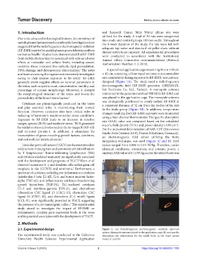
generally exerts negative effects on seminal parameters in designed (Figure 1A). The study used a radiofrequency
the testes, such as sperm count, concentration, motility, and electromagnetic field (RF-EMF) generator (GHZ2011X,
percentage of normal morphology. Moreover, it disrupts Set Electronic Co. Ltd., Turkey). A monopole antenna
the morphological structure of the testes and boosts the connected to the generator emitted 900 MHz RF-EMR and
permeability of the blood–testis barrier. 1 was placed in the application cage. The monopole antenna
was strategically positioned to evenly radiate RF-EMR at
Cytokines are physiologically produced in the testes a consistent distance of 32 cm from the bodies of the rats
and play essential roles in maintaining their normal in the study group (Figure 1B). In addition, temperature
function. However, cytokines also have critical roles in changes resulting from RF-EMR exposure were monitored
inducing inflammatory reactions under stress conditions. using a four-channel thermometer. The specific absorption
Exposure to RF-EMR leads to an increase in reactive rate (SAR) value was computed based on the calculated
oxygen species (ROS) and oxidative stress. ROS generated electric field density (V/m) and power density (mW/cm ).
2
2
by oxidative stress activates nuclear factor kappa B (NF-κB) For the numerical determination of SAR, CST (Microwave
and activator protein-1. In addition, it stimulates the Studio Suite [version 2018], Dassault Systèmes, Germany),
transcription of genes encoding growth factors, cytokines, an electromagnetic field solver utilizing the finite
and extracellular matrix proteins. 1 integration technique, was used (Figure 1C and D). SAR
Testicular germ cell tumors (TGCT) are the most prevalent values ranged from 0.008 to 0.14 W/kg. Therefore, under
solid tumors in young men and commonly exhibit infiltration identical conditions, orientation, and antenna power, a
3
by T lymphocytes. Tumor-infiltrating lymphocytes (TIL) uniform SAR value of 0.14 W/kg across the whole body was
and cytokine-mediated immunity are significantly associated
with the development and prognosis of TGCT. Klein et al. A B
4
observed numerous B, T, and dendritic cells within germ cell
4
neoplasia in situ (GCNIS) and seminoma. Furthermore, a
spectrum of cytokines, including pro-inflammatory cytokines
(interleukin 1 beta [IL-1β], IL-6, and tumor necrosis factor-
alpha [TNF-α]), anti-inflammatory cytokines (transforming
growth factor-beta [TGF-β1), Th1-mediated cytokines
(IL-2 and interferon-gamma [IFN-γ]), and chemokines
(chemokine CXC ligand 13 [CXCL-13], chemokine CXC
ligand 10 [CXCL-10], and chemokine [C-C motif] ligand C D
[CCL-5]), were significantly presented in TGCT, suggesting
4
the presence of a pro-tumorigenic milieu. This experimental
study aimed to investigate the impact of RF-EMR on
inflammatory cytokine gene expression levels in the testes
and its potential association with the development of TGCT.
2. Methods
2.1. Experimental design Figure 1. (A) Radiofrequency electromagnetic radiation exposure
system. Monopole antenna placed in the application cage (B) and specific
The experimental study was conducted at the Cukurova absorption rate distribution for the whole body in the voxel mouse
University Health Sciences Experimental Application model (C and D).
Volume 3 Issue 1 (2024) 2 https://doi.org/10.36922/td.1703