
Page 40 - TD-3-2
P. 40
Tumor Discovery MPTT post-chemotherapy
for 20 years but started growing rapidly over the past metastasis was detected during the 18-month follow-up
10 months, becoming painful, itchy, and spontaneously period.
bleeding. Thirteen years ago, the patient was diagnosed
with breast carcinoma, for which she underwent radical 3. Discussion
mastectomy with lymph node dissection, followed by The first case of trichilemmal cysts was reported by Wilson
radiotherapy, chemotherapy, and hormonotherapy. Jones in 1966. Trichilemmal cysts occur in 5% to 10% of
Unfortunately, access to the patient’s medical records was the population, with barely 2% progressing into PTTs.
3,6
not possible. The patient also reported a family history The term MPTT was first proposed by Headington in
7
of multiple pilar cysts and disclosed having four of them 1976 and later adopted by Saida et al. in 1983 to define
8
removed 3 years ago. There was no history of trauma or a PTT exhibiting malignant features such as infiltrative
chronic irritation. During the clinical examination, a firm, growth pattern, cytological atypia, atypical mitosis, and
nodular mass was identified on the parietal region of the lymph node metastasis. Despite being rare, MPTTs
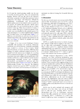
scalp, measuring 4 × 3 cm, with irregular borders, a hairless predominantly arise in the scalp region (90%), with 84%
and ulcerated surface, and telangiectasia (Figure 1). In of cases reported in mainly women aged between 40 and
addition, seven subcutaneous cysts were discovered in the 80 years. Other implicated locations include the face,
9,10
different areas of the scalp, ranging in size between 0.5 cm breast, back, chest, abdomen, buttocks, pubis, vulva, back
and 2 cm (Figure 2). Notably, no palpable neck lymph of hand, and wrist. 9,11
nodes were detected.
MPTTs are usually characterized as slowly enlarging,
A biopsy of the mass revealed histological features firm, painless nodular lesions ranging in size from 1 to
compatible with squamous cell carcinoma. Subsequent 30 cm, often with unremarkable cutaneous covering.
craniofacial and cervical-thoracic computed tomography These nodules persist for a significant duration before
scans revealed no evidence of bone invasion, lymph undergoing abrupt and rapid tumor evolution, marked by
node involvement, or distant metastasis. Following these pain, ulceration, bleeding, or purulent discharge, indicative
findings, the patient underwent surgical excision of the of malignant metamorphosis. 1,9,10,12-14 Cases of regional
parietal lesion with macroscopical margins of 1 cm, along lymph node metastasis have also been reported. 8,13-15
with the removal of the largest cyst. The final histological
examination depicted a malignant tumor proliferation with Although malignant degeneration is rare, MPTTs
lobulated architecture, coupled with focal stromal invasion commonly arise from preexisting trichilemmal cysts.
demonstrating increasing nuclear and cytoplasmic Alternatively, several authors have reported cases of MPTTs
16
pleomorphism, accompanied by numerous and often originating de novo, from organoid nevi or seborrheic
9
atypical mitotic figures (Figure 3A, B and C). In addition, keratosis. Many researchers have suggested a hypothetical
lymphovascular invasion was observed. The definitive malignant transformation pathway between trichilemmal
diagnosis rendered was a parietal MPTT associated with cysts and MPTTs progressing from the adenomatous
multiple trichilemmal cysts. Remarkably, no recurrence or stage of trichilemmal cysts to the epitheliomatous stage
of PTTs and eventually to the carcinomatous stage of
MPTTs. 3,4,9,10,12,17 As in our case, the commonly found
accompaniment of MPTTs with simple trichilemmal cysts
may support this theory, as does the coexistence of benign
and malignant areas within the same neoplasm. Trauma
9
and chronic inflammation are the most incriminated
factors in this degeneration. Only two cases of MPTT
associated with chemotherapy have been reported in the
literature; further research and reports are necessary to
4,5
establish a solid causality link between each of these factors
and MPTTs.
MPTTs may be easily confused with squamous cell
carcinoma since they share many clinical similitudes
and are both rare cases. Similarly, MPTTs must be
distinguished from PTTs or trichilemmal carcinoma
because they affect prognosis and treatment approach.
Figure 1. Nodular mass of the parietal scalp with irregular ulcerated Histologically, MPTT is described as a tumor that
borders, hairless surface, and telangiectasia. invades neighboring tissues accompanied by anaplasia,
Volume 3 Issue 2 (2024) 2 doi: 10.36922/td.2344