
Page 41 - TD-3-2
P. 41
Tumor Discovery MPTT post-chemotherapy
Figure 2. Mapping of the patient’s nodular scalp lesions.
A B C
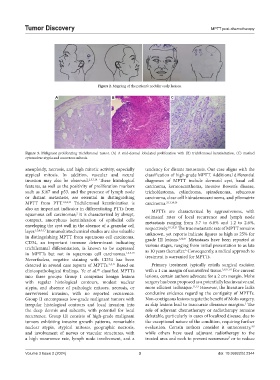
Figure 3. Malignant proliferating trichilemmal tumor. (A) A mid-dermal lobulated proliferation with (B) trichilemmal keratinization, (C) marked
cytonuclear atypia and numerous mitosis.
aneuploidy, necrosis, and high mitotic activity, especially tendency for distant metastasis. Our case aligns with the
atypical mitosis. In addition, vascular and neural classification of high-grade MPTT. Additional differential
invasion may also be observed. 2,5,7,18 These histological diagnoses of MPTT include dermoid cyst, basal cell
features, as well as the positivity of proliferation markers carcinoma, keratoacanthoma, invasive Bowen’s disease,
such as Ki67 and p53, and the presence of lymph node trichoblastoma, cylindroma, spiradenoma, sebaceous
or distant metastasis, are essential in distinguishing carcinoma, clear cell hidradenocarcinoma, and pilomatrix
MPTT from PTT. 1,2,4,19 Trichilemmal keratinization is carcinoma. 3,11,14,16
also an important indicator in differentiating PTTs from MPTTs are characterized by aggressiveness, with
squamous cell carcinomas; it is characterized by abrupt, estimated rates of local recurrence and lymph node
2
compact, amorphous keratinization of epithelial cells metastasis ranging from 3.7 to 6.6% and 1.2 to 2.6%,
enveloping the cyst wall in the absence of a granular cell respectively. 10,13,21 The true metastatic rate of MPTT remains
layer. 1,2,4,9,13 Immunohistochemical studies are also valuable unknown, yet reports indicate figures as high as 25% for
in distinguishing MPTT from squamous cell carcinoma. grade III lesions. 3,20,21 Metastases have been reported at
CD34, an important immune determinant indicating various stages, ranging from initial presentation to as late
trichilemmal differentiation, is known to be expressed as 10 years thereafter. Consequently, a radical approach to
6
in MPTTs but not in squamous cell carcinomas. 1,2,4,19 treatment is warranted for MPTTs.
Nevertheless, negative staining with CD34 has been
detected in several case reports of MPTTs. 2,4,17 Based on Primary treatment typically entails surgical excision
clinicopathological findings, Ye et al. classified MPTTs with a 1 cm margin of uninvolved tissue. 3,9,11,16 For current
20
into three groups: Group I comprises benign lesions lesions, certain authors advocate for a 2 cm margin. Mohs
with regular histological contours, modest nuclear surgery has been proposed as a potentially less invasive and
atypia, and absence of pathologic mitoses, necrosis, or more efficient technique. 22,23 However, the literature lacks
nerve/vessel invasion, with no reported recurrence. conclusive evidence regarding the contiguity of MPTTs.
Group II encompasses low-grade malignant tumors with Non-contiguous lesions negate the benefit of Mohs surgery,
3
irregular histological contours and local invasion into as skip lesions lead to inaccurate clearance margins. The
the deep dermis and subcutis, with potential for local role of adjuvant chemotherapy or radiotherapy remains
recurrence. Group III consists of high-grade malignant debatable, particularly in cases of localized disease, due to
tumors exhibiting invasive growth patterns, remarkable the exceptional nature of the condition, requiring further
nuclear atypia, atypical mitoses, geographic necrosis, evaluation. Certain authors consider it unnecessary,
3,9
and involvement of nerves or vascular structures, with while others have used adjuvant radiotherapy to the
a high recurrence rate, lymph node involvement, and a treated area and neck to prevent recurrence or to reduce
7
Volume 3 Issue 2 (2024) 3 doi: 10.36922/td.2344