
Page 83 - TD-4-3
P. 83
Tumor Discovery Mg-28-A theoretical novel strategy in cancer therapy
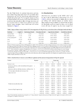
Van der Waals bonds. In contrast, beta-minus particles, 3.3. Absorbed dose
due to their long range and lowest LET, primarily affect 26
hydrophobic and Van der Waals bonds and produce free Absorbed dose calculations by the MIRD code, with
radicals along their path. The therapeutic impact can thus 0.1 ng of Mg-28 administered intravenously over 21 h
be characterized by the focused molecular-level action of (Table 4), showed the potential for highly effective tumor
recoil ions, the intracellular effect of Auger electrons, and cell killing with minimal impact on healthy tissues.
the extended influence of beta-minus particles up to 6mm Three regimens were modeled: (a) Whole-body cell dose,
beyond tumor margins. These distinctions are further (b) Mg-uptake coefficient-based cell dose, and (c) direct
illustrated in Figure 1. tumor cell dose. In regimen (a), the doses ranged from
Table 3. Impact of linear energy transfer (LET) from magnesium‑28 (Mg‑28) decay on the dissociation of biological molecular bonds
Bond type Length (Å) Bond energy (kJ/mol) Beta minus (kJ/mol) Auger electron (kJ/mol) Recoiled ions (kJ/mol)
Van der Waals 3.0–4.0 ~ 2.4–4 7.08–34.84 311.81–612.41 38.12–81.08
Hydrogen bond 1.5–2.0 ~4–40 3.54–17.42 155.91–306.21 19.06–40.54
P–O (in ATP/ADP) * ~1.60 ~30.5 2.83–13.94 124.72–244.96 15.25–32.43
28
Ionic 2.0–4.0 ~20–200 7.08–34.84 311.81–612.41 38.12–81.08
N–N ~1.45 ~163 2.57–12.63 112.88–222.00 13.82–29.39
S–S 2.05 ~250 3.64–17.82 156.31–381.20 19.46–41.54
Notes: (1) *Bond energy released when ATP hydrolyzes to ADP; 1eV=96.485 kJ/mol. (2) Van der Waals interactions (3.0–4.0 Å) are weak but critical
for stabilizing protein side chains and stacked DNA bases. Hydrogen bonds (1.5–2.0 Å) maintain DNA base pairing (A=T, G≡C) and play a key role
in enzyme-substrate interactions. Ionic (Coulombic) interactions (2.0–4.0 Å) are prominent between oppositely charged side chains of proteins and
in ATP–Mg coordination complexes. Phosphodiester (P–O) bonds (~1.60 Å) form the DNA/RNA backbone and are targets for strand cleavage
2+
during radiation damage. Disulfide bridges (S–S) (~2.05 Å) stabilize the tertiary/quaternary structure of many enzymes, particularly in oxidative
environments. Nitrogen–nitrogen (N–N) bonds (~1.45 Å) are present in some nucleotide base pairs and enzyme cofactors (e.g., flavins). The LET
values from Mg-28 decay are sufficient to break all the above interactions, directly impairing nucleic acid integrity and enzymatic function essential to
cellular integrity, especially in cancer cells.
Table 4. Absorbed dose to tumors across different regimens and the amount of magnesium‑28 (Mg‑28) required
Content Regimen Cancer type
T 0 T 1 T 2 T 3 T 4 T 5
Number of cells 3.28E+04 5.00E+06 5.00E+07 5.00E+08 5.00E+09 5.00E+11
Intravenous cell dose, uniformly distributed throughout a 1.01E-11 1.55E-09 1.55E-08 1.55E-07 1.55E-06 1.55E-04
the body (Gy)
Intravenous cell dose with Mg absorption coefficient (Gy) b 1.83E-09 3.46E-06 1.09E-04 3.46E-03 1.09E-01 1.09E+02
Cell dose injected directly into the tumor (Gy) c 3.04E+04 2.23E+02 2.23E+01 2.23E+00 2.23E-01 2.23E-02
Tumor dose (Gy) a 3.31E-07 7.75E-03 7.75E-01 7.75E+01 7.75E+03 7.75E+07
b 6.00E-05 11.15E+00 11.15E+03 11.15E+05 11.15E+08 11.15E+13
c 9.97E+08 38.75E+08 38.75E+08 38.75E+08 38.75E+08 38.75E+09
d 6.01E+01 6.01E+01 6.01E+01 6.01E+01 6.01E+01 6.01E+01
e 3.12E+02 3.12E+02 3.12E+02 3.12E+02 3.12E+02 3.12E+02
f 4.15E+02 4.15E+02 4.15E+02 4.15E+02 4.15E+02 4.15E+02
Whole-body absorbed dose (Gy) d 3.52E-01 2.03E-02 8.91E-03 2.82E-03 8.91E-04 8.91E-05
e 1.72E+00 9.81E-02 4.42E-02 1.44E-02 4.42E-03 4.42E-04
f 2.33E+00 1.32E-01 5.92E-02 1.92E-02 5.92E-03 5.92E-04
Amount of Mg-28 required (ng) d 9.31E-01 5.33E-02 2.42E-02 7.51E-03 2.44E-03 2.44E-04
e 4.62E+00 2.63E-01 1.22E-01 3.71E-02 1.22E-02 1.22E-03
f 6.21E+00 3.51E-01 1.63E-01 5.04E-02 1.63E-02 1.63E-03
Note: All absorbed dose calculations are based on a 21-h decay duration. Regimen (d) assumes 62 Mg-28 ions/cell, regimen (e) assumes 300 ions/cell,
and regimen (f) assumes 400 ions/cell.
Volume 4 Issue 3 (2025) 75 doi: 10.36922/TD025070010