
Page 49 - manuscript_ijb05590
P. 49
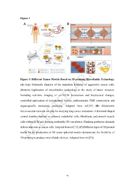
Figure 3


                   Figure 3: Different Tumor Models Based on 3D printing Microfluidic Technology.

                   (A)  (top)  Schematic  diagram  of  the  migration  behavior  of  aggressive  cancer  cells.

                   (Bottom) Application  of  microfluidics  technology  in  the  study  of  tumor  invasion.

                   Including  real-time  imaging  of  cell-ECM  interactions  and  biophysical  changes,
                   controlled application of promigratory factors, pathomimetic TME construction and


                   organ-specific  metastasis  modeling.  Adapted  from  ref.[69]  (B)  Biomimetic
                   microvascular network-on-chip for studying lung cancer metastasis. A diamond-shaped

                   central chamber harbors co-cultured endothelial cells, fibroblasts, and smooth muscle

                   cells within ECM gel, forming perfusable 3D vasculature. Flanking perfusion channels

                   deliver nutrients or cancer cells. Adapted from ref.[72] (C) Different types of 3D printed

                   molds for the production of 3D tumor spheroid models demonstrate the flexibility of

                   3D printing to produce microfluidic devices. Adapted from ref.[73]


                                                            48