
Page 50 - manuscript_ijb05590
P. 50
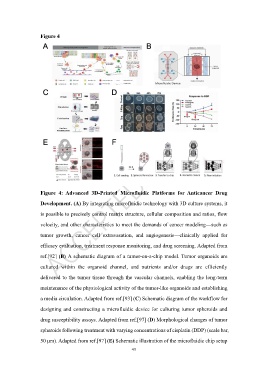
Figure 4


                   Figure  4:  Advanced  3D-Printed  Microfluidic  Platforms  for  Anticancer  Drug

                   Development. (A) By integrating microfluidic technology with 3D culture systems, it
                   is possible to precisely control matrix structure, cellular composition and ratios, flow

                   velocity, and other characteristics to meet the demands of cancer modeling—such as

                   tumor  growth,  cancer  cell  extravasation,  and  angiogenesis—clinically  applied  for

                   efficacy evaluation, treatment response monitoring, and drug screening. Adapted from

                   ref.[92] (B) A schematic diagram of a tumor-on-a-chip model. Tumor organoids are

                   cultured  within  the  organoid  channel,  and  nutrients  and/or  drugs  are  efficiently

                   delivered to the tumor tissue through the vascular channels, enabling the long-term

                   maintenance of the physiological activity of the tumor-like organoids and establishing

                   a media circulation. Adapted from ref.[93] (C) Schematic diagram of the workflow for

                   designing and constructing a microfluidic device for culturing tumor spheroids and

                   drug susceptibility assays. Adapted from ref.[97] (D) Morphological changes of tumor

                   spheroids following treatment with varying concentrations of cisplatin (DDP) (scale bar,

                   50 μm). Adapted from ref.[97] (E) Schematic illustration of the microfluidic chip setup
                                                            49