
Page 124 - AIH-2-3
P. 124
Artificial Intelligence in Health Opportunities for AI-based arrhythmia screening
Table 2. A prospective spectrum of cardiac rhythm
diagnostics and screening that can be quickly and
automatically derived from an electrocardiogram
Number Content
1 1 degree atrioventricular block
st
2 Atrial fibrillation
3 Atrial flutter
4 Bradycardia
5 Complete the right bundle branch block
6 Hypertrophic cardiomyopathy (also known as left
ventricular hypertrophy)
7 Incomplete right bundle branch block
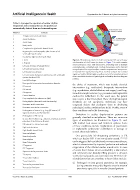
8 J-point Figure 6. The infarcted volume in the left ventricular (LV) wall will cause
9 J-60 point a deformation of the R wave (as shown in Figure 7) in each recursive
10 Left-axis deviation (Purkinje fibers) electrocardiogram cycle. This deformation, presenting itself as multiple
conjoined peaks, a stretched peak, or other phenomena, can be detected
11 Left anterior fascicular block using adaptive filter signal analysis, wavelet computational comparison,
12 Left bundle branch block or matched filter signal processing. These methods compare the recorded
13 Left ventricular dysfunction (defined as a left ventricular signal to a healthy QRS template, as well as several other templates derived
ejection fraction≤35%) from a worldwide database of pathological and healthy electrocardiogram
patterns.
14 Low QRS voltages
15 Non-specific Intraventricular conduction disorder the choice of treatments, which may include chemical
16 PP interval interventions (e.g., medication), therapeutic interventions
17 PR interval (e.g., cryoablation, alcohol ablation, and surgery), and long-
18 PR segment term device implementation (e.g., pacemaker and implantable
19 P wave duration cardioverter defibrillator. In the worst case, the patient
20 P top amplitude (in reference to QRS) may require a heart transplant. These electrophysiological
21 Pacing rhythm (sinoatrial node functionality) deviations are not age-specific. Individuals may have
22 Premature atrial contraction congenital defects that predispose them to developing
23 Premature ventricular contraction pathological cardiac conditions later in life. Notably, some of
24 Prolongation of interval (e.g., long QT interval, ST these conditions can be life-threatening.
segment duration, and PQ interval)
Deviations in cardiac depolarization patterns are
25 Prolongation of PR interval generally classified as arrhythmias. There are numerous
26 Q wave abnormalities (e.g., duration, amplitude, deletion/ types of arrhythmia (as illustrated in Figure 7), each
reduction of the follow-on R wave.)
27 RR interval (i.e., derive heart rate) with distinct root causes and treatment options. Some
of these conditions include implantation of pacemakers
28 Right-axis deviation
29 Right bundle branch block or implantable cardioverter defibrillators to manage or
correct abnormal rhythms.
30 Sinus arrhythmia
31 Sinus bradycardia One particularly life-threatening arrhythmia is VF,
32 Sinus rhythm which can lead to SCD if not immediately treated. 4,47-50 VF
33 Sinus Tachycardia can be triggered by heart block and coronary artery disease,
34 ST segment which is characterized by impaired perfusion and reduced
35 ST–T segment oxygenation of the affected cardiac muscle cells. In cases
36 Supraventricular premature beats of severe heart failure, where contractility is diminished,
and intraventricular diastolic filling pressure is elevated,
37 TP interval ventricular tachycardia can deteriorate into VF. Other life-
38 T wave abnormalities related factors that can contribute to heart failure include:
39 T wave inversion (i) Prolonged or deep general anesthesia, for example,
40 Ventricular premature beats during extensive myocardial hypoxia or at the onset of
41 Ventricular fibrillation anesthesia, especially in severely diseased patients
Volume 2 Issue 3 (2025) 118 doi: 10.36922/aih.8468