
Page 117 - AN-3-3
P. 117
Advanced Neurology Evaluating plausibility of thalamic model
Clinical and experimental studies demonstrate that the stabilized images not only depict a phenomenon localized
thalamus controls the alternation and level of wakefulness to the eye but also involve higher brain areas.
and sleep states, 35-37 with lesions in the non-specific Llinás et al. initial proposal on resonant columns was
22
pathways 38,39 often resulting in loss of consciousness. pioneering in attempting to computationally understand
Cortical layer V pyramidal neurons, which have extensive the functioning of thalamocortical networks. Subsequently,
connections with the thalamus, are central to this theory, various models emerged, exploring the different operational
implying that any cortical processing excluding these contexts of this circuit. 41-44 Han et al. developed a
45
neurons will be unconscious. Stimulation of neurons in computational model based on multi-scale recurrent neural
the centrolateral nucleus of the thalamus in anesthetized
primates restores the wakeful state, suggesting clinical networks with synaptic depression to elucidate novelty
applications for alleviating consciousness disorders. 36,37 detection in the whisker-related region (barrel cortex) of
the rat somatosensory thalamocortical circuit. Meanwhile,
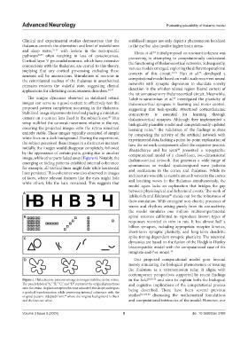
The unique dynamics observed in stabilized retinal Lakshminarasimhan et al. investigated the plasticity of
46
images can serve as a good context to effectively test the thalamocortical synapses in learning and motor control,
proposed pattern completion occurring in the thalamus. suggesting that task-specific structured corticothalamic
Stabilized image experiments involved placing a miniature connectivity is essential for learning through
camera on a contact lens fixed in the subject’s eye. This thalamocortical synapses. Although they implemented a
40
setup nullified the camera’s movement relative to the eye, biologically plausible model and computationally updated
ensuring the projected images onto the retina remained learning rules, the validation of the findings is done
47
entirely stable. These images typically consisted of simple by comparing the activity of the artificial network with
white lines on a dark background. During the experiment, experimental data, lacking a point-by-point explanation of
the subject perceived these images in a distinctive manner: how the network components affect the cognitive process.
initially, the images would disappear completely, followed Bhattacharya and his team presented a topographic
48
by the appearance of certain parts, giving rise to another computational model of a closed-loop, two-dimensional
image, while other parts faded away (Figure 4). Notably, the thalamocortical network that generates a wide range of
emerging or fading patterns exhibited internal coherence; spontaneous or evoked spatiotemporal wave patterns
for example, all vertical lines might fade while horizontal and oscillations in the cortex and thalamus. While its
lines persisted. This coherence was also observed in images architecture was able to sustain smooth waves in the cortex
of faces, where relevant features like the eyes might fade and lurching waves in the thalamus simultaneously, the
while others, like the hair, remained. This suggests that
model again lacks an explanation that bridges the gap
between physiological and behavioral events. The work of
A
Izhikevich and Edelman stands out for the robustness of
20
their simulation. With emergent non-chaotic processes of
waves and rhythms arising purely from its connectivity,
the model simulates one million multicompartmental
B spinal neurons calibrated to reproduce known types of
responses recorded in vitro in rats. It has almost half a
billion synapses, including appropriate receptor kinetics,
short-term synaptic plasticity, and long-term dendritic
C spike-timing-dependent synaptic plasticity. The neuronal
dynamics are based on the fusion of the Hodgkin-Huxley
biocompatible model with the computational ease of the
integrate-and-fire model. 49
D Our proposed computational model goes beyond
merely simulating the biological phenomenon or treating
the thalamus as a retransmission relay. It aligns with
contemporary perspectives supported by recent findings
Figure 4. Hallucinatory patterns emerge as images stabilize on the retina. in the field 28,50-52 and aims to explain both the biological
The panels labeled “A,” “B,” “C,” and “D” represent the original projections and cognitive implications of the computational process
onto the retina. As photoreceptors become saturated, the shape undergoes being described. There have been several previous
a gradual transformation while preserving internal coherence with the 8,11,53,54
original pattern. Adapted from, where the original background is black studies discussing the mathematical foundations
40
and the lines are white. and computational intricacies of the model. However, our
Volume 3 Issue 3 (2024) 5 doi: 10.36922/an.3188