
Page 62 - ARNM-1-1
P. 62
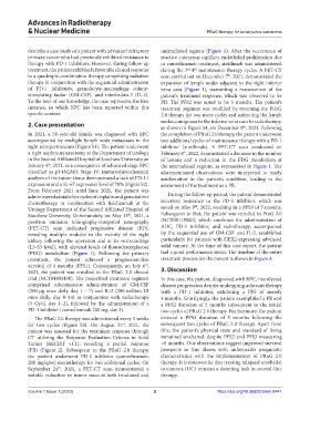
Advances in Radiotherapy
            & Nuclear Medicine                                                  PRaG therapy for renal pelvis carcinoma


            describe a case study of a patient with advanced refractory   unirradiated regions (Figure  1). After the occurrence of
            primary cancer who had previously exhibited resistance to   reactive cutaneous capillary endothelial proliferation due
            therapy with PD-1 inhibitors. However, during follow-up   to camrelizumab treatment, sintilimab was administered
            treatment, the patient exhibited a favorable clinical response   during the 3 -4  maintenance therapy cycles. A PET-CT
                                                                            th
                                                                         rd
            to a quadruple-combination therapy comprising radiation   scan carried out on December 7 , 2021, demonstrated the
                                                                                         th
            therapy in conjunction with the sequential administration   expansion of  lymph  nodes  adjacent  to the right  inferior
            of PD-1 inhibitors, granulocyte-macrophage colony-  vena cava (Figure  1), warranting a reassessment of the
            stimulating factor (GM-CSF), and interleukin-2 (IL-2).   patient’s treatment response, which was observed to be
            To the best of our knowledge, the case represents the first   PD. The PFS2 was noted to be 5  months. The patient’s
            instance, in which RPC has been reported within this   treatment regimen was modified by resuming the PRaG
            specific context.                                  2.0 therapy for two more cycles and subjecting the lymph

            2. Case presentation                               nodes contiguous to the inferior vena cava to radiotherapy,
                                                               as shown in Figure S4, on December 9 , 2021. Following
                                                                                              th
            In 2021, a 58-year-old female was diagnosed with RPC   the completion of PRaG 2.0 therapy, the patient underwent
            accompanied  by  multiple  lymph  node  metastases  in  the   two additional cycles of maintenance therapy with a PD-1
            right retroperitoneum (Figure S1). The patient underwent   inhibitor (sintilimab). A  PET-CT scan conducted on
            a right nephroureterectomy at the Department of Urology   February 8 , 2022, demonstrated a decrease in the number
                                                                       th
            in the Second Affiliated Hospital of Soochow University on   of lesions and a reduction in the FDG metabolism at
            January 4 , 2021, as a consequence of advanced stage RPC   the unirradiated regions, as represented in Figure 1. The
                   th
            classified as pT4N2M0, Stage IV. Immunohistochemical   aforementioned  observations  were  interpreted  to  imply
            analysis of the tumor tissue demonstrated a lack of PD-L1   amelioration in the patient’s condition, leading to the
            expression and a Ki-67 expression level of 70% (Figure S2).   assessment of the treatment as a PR.
            From February 2021 until June 2021, the patient was
            administered a total of six cycles of cisplatin and gemcitabine   During the follow-up period, the patient demonstrated
            chemotherapy in combination with tislelizumab at the   recurrent resistance to the PD-1 inhibitor, which was
                                                                            th
            Urology Department of the Second Affiliated Hospital of   noted on May 9 , 2022, resulting in a PFS3 of 5 months.
            Soochow University. Unfortunately, on May 13 , 2021, a   Subsequent to that, the patient was enrolled in PraG 3.0
                                                  th
            positron emission tomography-computed tomography   (NCT05115500), which combines the administration of
            (PET-CT) scan indicated progressive disease (PD),   ADC, PD-1 inhibitor, and radiotherapy, accompanied
            revealing multiple nodules in the vicinity of the right   by the sequential use of GM-CSF and IL-2, established
            kidney  following  the  operation  and  at  its  surroundings   particularly for patients with HER2-expressing advanced
            (L5-S3 level), with elevated levels of fluorodeoxyglucose   solid tumors. At the time of this case report, the patient
            (FDG) metabolism (Figure  1). Following the primary   had a good performance status. The timeline of the entire
            treatment, the patient achieved a progression-free   treatment process for the patient is shown in Figure 3.
            survival of 4  months (PFS1). Consequently, on July 6 ,
                                                         th
            2021, the patient was enrolled in the PRaG 2.0 clinical   3. Discussion
            trial (NCT04892498). The prescribed treatment regimen   In this case, the patient, diagnosed with RPC, manifested
            comprised subcutaneous administration of GM-CSF    disease progression despite undergoing adjuvant therapy
            (200 µg once daily, day 1 – 7) and IL-2 (200 million UI   with a PD-1 inhibitor, exhibiting a PFS of merely
            once daily, day 8–14)  in conjunction with radiotherapy   4 months. Gratifyingly, the patient exemplified a PR and
            (5  Gy/d, day 1–2), followed by the administration of a   a PFS2 duration of 5  months subsequent to the initial
            PD-1 inhibitor (camrelizumab 200 mg, day 3).       two cycles of PRaG 2.0 therapy. Furthermore, the patient
              The PRaG 2.0 therapy was administered every 3 weeks   evinced a PFS3 duration of 5  months following the
                                                st
            for two cycles (Figure S3). On August 31 , 2021, the   subsequent two cycles of PRaG 2.0 therapy. Apart from
            patient was assessed for the treatment response through   this, the patient’s physical state and standard of living
            CT utilizing the Response Evaluation Criteria in Solid   remained unaltered, despite PFS2 and PFS3 measuring
            Tumor (RECIST v1.1), revealing a partial response   <6 months. Our observations suggest improved survival
            (PR) (Figure  2). Subsequent to the PRaG 2.0 therapy,   prospects in this illness with unfavorable prognostic
            the patient underwent PD-1 inhibitor (camrelizumab,   characteristics with the implementation of PRaG 2.0
            200 mg/q3w) monotherapy for two additional cycles. On   therapy. It is noteworthy that treating relapsed urothelial
            September 24 , 2021, a PET-CT scan demonstrated a   carcinoma (UC) remains a daunting task in second-line
                       th
            notable reduction in tumor mass at both irradiated and   therapy.

            Volume 1 Issue 1 (2023)                         2                       https://doi.org/10.36922/arnm.0441