
Page 73 - ARNM-3-1
P. 73
Advances in Radiotherapy
& Nuclear Medicine Aspirin’s protective effect on RISI
A B C
D E
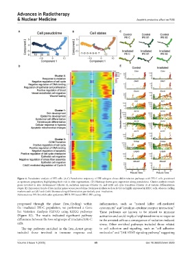
Figure 4. Pseudotime analysis of IFE cells. (A-C) Pseudotime trajectory of IFE subtypes shows differentiation pathways with IFE C cells positioned
as upstream progenitors, highlighting their role in skin regeneration. (D) Heatmap shows gene expression along pseudotime. Cluster analysis reveals
genes involved in skin development (Cluster 1), radiation response (Cluster 2), and G2M cell cycle transition (Cluster 3) at various differentiation
stages. (E) Expression trends of key marker genes across pseudotime. Stemness markers such as Krt14 are highly expressed in IFE C cells, whereas cycling
markers such as Cdk1 and Ccnb1 decrease along differentiation, particularly post-irradiation.
Abbreviations: IFE: Interfollicular epidermis; IFE B: IFE basal; IFE C: IFE cycling.
progressed through the phase (Irra_Cycling) within inflammation, such as “natural killer cell-mediated
the irradiated IFE-C population, we performed a Gene cytotoxicity” and “cytokine-cytokine receptor interaction.”
Set Variation Analysis (GSVA) using KEGG pathways These pathways are known to be related to immune
(Figure S2). The results indicated significant pathway activation and could imply a heightened immune response
differences between the two subgroups of irradiated IFE-C in the arrested cells as a consequence of radiation-induced
cells. stress. Other enriched pathways included those related
The top pathways enriched in the Irra_Arrest group to cell adhesion and signaling, such as “cell adhesion
included those involved in immune response and molecules” and “JAK-STAT signaling pathway,” suggesting
Volume 3 Issue 1 (2025) 65 doi: 10.36922/arnm.5829