
Page 72 - ARNM-3-1
P. 72
Advances in Radiotherapy
& Nuclear Medicine Aspirin’s protective effect on RISI
A B E F
G H
C D
I J
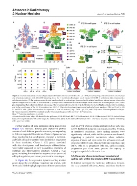
Figure 3. Detailed characterization and subtype analysis of irradiated versus control skin cells. (A) t-SNE plot comparing cell distributions in control (blue)
and irradiated conditions (red). (B) t-SNE clustering shows the 13 identified cell subtypes, which include IFE B, IFE D, and others, after comprehensive
cell characterization. (C) Bar plot represents the total number of cells in each subtype for control and irradiated conditions, with an increase observed in
specific subtypes such as IFE B1 in irradiated skin. (D) Proportional distribution of each cell subtype across control and irradiated groups. (E-G) t-SNE
plots highlighting the localization of IFE B cells among other epidermal cell types, the cell cycle distribution (G1, S, G2/M phases) in the IFE B population,
and further stratification of the IFE B1 population into IFE C. (H) Density plots show the expression levels of cell cycle regulators Cdk1 and Ccnb1 in IFE
B1 and IFE C cells. (I) Dot plot visualizes the expression of stem cell markers (Lrig1, Krt14, Lgr5, Gli1, and Cd34) across different epidermal cell types.
(J) Higher expression of Krt14 in irradiated samples whereas cycling genes Cdk1 and Ccnb1 show a decrease.
Note: ****indicates significance at p<0.001.
Abbreviations: IB: Inner bulge; IFE: Interfollicular epidermis; IFE B: IFE basal; IFE D: IFE differentiated; IFE K: IFE keratinized; INFU B: Infundibulum
basal; LH: Langerhans cells; OB: Outer bulge; SG: Subcutaneous gland; SCMs: Stem cell markers; t-SNE: t-Distributed stochastic neighbor embedding;
uHF: upper-hair follicle.
Further analysis of gene expression along pseudotime such as Krt14, whereas cycling markers such as Cdk1 and
(Figure 4D) indicated distinct gene expression profiles Ccnb1 decreased along the differentiation path. Notably,
associated with different pseudotime states, corresponding in irradiated conditions, these cycling markers were
to different stages of differentiation. Clusters of genes such as significantly reduced, while Krt14 expression was elevated,
those involved in skin development, response to radiation, suggesting a potential mechanism where radiation
and G2M cell cycle transition were identified as key players exposure induces cell cycle arrest but enhances stem cell
in these transitions. Cluster 1 (C1) genes, associated properties of IFE C cells. This supports our hypothesis that
with skin development and keratinocyte differentiation, IFE C cells act as progenitor cells and, upon successful
were highly expressed in early pseudotime, indicative of progression past G2M arrest, possess the capacity to
stemness and differentiation capability. Cluster 2 (C2) repopulate epithelial cells in the skin.
genes, including those related to response to radiation and
cell cycle regulation, were more prominent in later stages. 3.5. Molecular characterization of arrested and
cycling cells within the irradiated IFE-C population
In Figure 4E, the expression dynamics of key marker
genes along the pseudotime trajectory are shown, with To further investigate the molecular differences between
IFE C cells exhibiting high expression of stemness markers the G2M-arrested cells (Irra_Arrest) and those that have
Volume 3 Issue 1 (2025) 64 doi: 10.36922/arnm.5829