
Page 13 - BH-1-1
P. 13
Brain & Heart Application of the neural networks surgery
to investigate the interaction of different brain regions and middle frontal gyrus) involved in the integration
functionally or structurally . With the help of the graph of information in semantic processing (Figure 3) .
[43]
[6]
theory to analyze data from medical imaging, brain This model directs the focus onto hub protection to
networks can be described as a combination of nodes achieve cognitive maintenance following neurosurgical
(i.e., brain regions) connected by edges (i.e., white matter intervention. Through further investigations, the influence
tracts) . In brain networks, the majority of nodes have few of brain lesions on brain network topology has been
[5]
connections, while the remaining nodes, which are known identified. Yuan et al. have contributed a lesion model
as hubs, have many connections . Through the analysis to explore this issue and found that the damage to brain
[44]
of both structural and FC data, studies have revealed network hubs causes two different directions of change in
that these brain hubs are mainly located at the posterior network topology, one being more integrated (global) and
cingulate cortex/precuneus, medial prefrontal cortex, and the other being more segregated (local). The result further
lateral temporal and parietal cortices, among which most confirms the significant role of hubs in brain networks .
[51]
of these regions are considered as parts of the DMN, which
plays a significant role in the resting state [45-47] . The previous Previous studies have shown that neurosurgery has
studies have shown compelling evidence to validate the been the primary focus in the treatment of nervous
aforementioned theory. Based on resting-state FC MRI system diseases. Based on three data sets of fMRI and 127
defined hubs, Power et al. used two methods (identifying participants, Buckner et al. gained a consensus estimate
network nodes that participate in multiple sub-networks of cortical hubs; PET amyloid imaging in AD patients
of the brain, and identifying the spatial locations where compared with older controls showed high amyloid-β
several systems are represented within a small volume) to deposition in the locations of cortical hubs, consistent
[52]
confirm the previously accepted hubs in brain networks with the accepted hubs . This finding suggests that brain
and considered novel brain regions in both methods. Their network hubs may have an association with cognitive
research supports the earlier finding and identifies putative deficits. Aben et al. have developed a lesion impact score
hubs in brain networks . According to another study, that integrates information on infarct size with healthy
[48]
network hubs play a critical role in information transfer brain network topology to estimate the damage to network
during resting and task states by combining with regional hubs; the research has verified that this scoring system
cerebral blood flow (rCBF) and functional connectivity can predict the cognitive recovery of patients based on
[43]
strength (FCS). During resting state, FCS has high spatial the damage of hubs in brain networks . This evidence
correlation with rCBF; this correlation is stronger in the opens a new insight into the treatment of cerebrovascular
DMN and executive control network than in the visual disease; during surgery, the hubs need to be protected
and sensorimotor networks, having a connection-distance to achieve the purpose of cognitive function protection;
dependent relationship. However, during task state, the after surgery, the focal hemorrhage needs to be estimated
indices are related to task load. Meanwhile, rCBF and accurately; and pre-symptomatic intervention must be
metabolism are positively associated with either structural confirmed. Based on the topological feature of brain
or functional hubs. From the studies above, it can be networks, there is a huge possibility for neural function
concluded that there is a tight association between blood recovery if the network hubs are not damaged in surgery.
supply and brain functional topology during both rest and
task states, and the studies also indicate the importance of
functional hubs in brain networks [49,50] .
Since we know that hubs are vital in maintaining whole-
brain network stability, the primary task now is to identify
specific hubs in certain behavior. By combing with meta-
analyses and the graph-theoretic approach, a tri-network
model in human semantic processing has been proposed
by Xu et al., who have found that the semantic system is
topologically segregated into three brain modules: DMN,
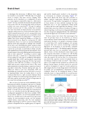
left perisylvian network, and left frontoparietal network. Figure 3. Three modules of semantic system. LFPN, DMN, and PSN
These three modules serve as the multimodal experiential serve as semantic control system, multimodal experiential system, and
system, the non-experiential system, and the semantic language-supported system, respectively. AG, angular gyrus; ATL,
control system, respectively, with five hubs (posterior anterior temporal lobe; pMTG, posterior middle temporal gyrus; pIPS,
posterior intraparietal sulcus; SFG/MFG, superior and middle frontal
middle temporal gyrus [pMTG], anterior temporal lobe, gyrus; LFN, left frontoparietal network; DMN, default mode network;
posterior intraparietal sulcus, angular gyrus, and superior PSN, perisylvian network.
Volume 1 Issue 1 (2023) 5 https://doi.org/10.36922/bh.v1i1.223