
Page 18 - BH-2-1
P. 18
Brain & Heart Pictorial rendition pulmonary stenosis
vessels. In 1967, Porstmann et al. described percutaneous A B
10
occlusion of patent ductus arteriosus (PDA). Shortly
11
thereafter, Rashkind and Cuaso developed different PDA
occluding devices. In 1976, King et al. introduced a
12
device to close atrial septal defects (ASDs). Subsequently,
13
Rashkind and Cuaso designed a different ASD occluding
device. In 1964, Dotter and Judkins proposed the concept
14
of stents. The introduction of the spiral coil-spring device
1
by Dotter and stainless steel mesh stents by Palmaz et al. 16
15
followed. The author utilized these devices and subsequently
developed transcatheter techniques during his academic
practice over the last four decades. Prospective data
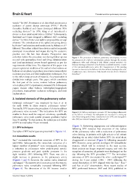
collection before the procedure and during follow-up was Figure 1. The procedure of balloon pulmonary valvuloplasty involves
secured with appropriate Food and Drug Administration the placement of a balloon valvuloplasty catheter through the stenotic
and local institutional review board approvals as per the pulmonary valve and inflating it with diluted contrast material. (A)
requirements of that time. The objective of this paper is to Balloon waisting is observed as the balloon is inflated (arrows), a result
present a pictorial rendition of the author’s observations on of the narrowed pulmonary valve. (B) Disappearance of the waisting
(arrows) is observed as the balloon is further inflated, leading to the relief
balloon valvuloplasty/angioplasty procedures, transcatheter of pulmonary valve obstruction. Only lateral views are shown. Modified
occlusion practices, and stent implantation techniques. Due from Rao. 19
to the voluminous amount of material, the presentation is
divided into multiple parts. This paper, which constitutes A B
the first part of the series, reviews balloon pulmonary
valvuloplasty (BPV) of pulmonary stenosis (PS). Subsequent
papers discuss other balloon valvuloplasty/angioplasty
procedures, transcatheter occlusion techniques, and stent
implantations.
2. Isolated stenosis of the pulmonary valve
Grüntzig’s technique was employed by Kan et al. in
2-5
the early 1980s to dilate stenotic pulmonary valves.
6
Eventually, BPV became the procedure of choice to address
pulmonary valve stenosis. The indications for BPV are Figure 2. (A) Balloon waisting in a neonate. (B) The waisting is eliminated
17
similar to those used for surgical valvotomy, specifically as the balloon is inflated. The radiograms are recorded in a sitting-up
pulmonary valve peak systolic pressure gradients higher view. Descending aorta (DAo), endotracheal tube (ET), and nasogastric
than 50 mmHg. In this section, the technique and results tube (NG) are labeled. Adopted from Rao et al. 20
18
of BPV to treat valvar PS are reviewed.
(Figure 7). Reviewing angiograms and echocardiograms
2.1. BPV techniques
following BPV revealed free excursion of the leaflets
Examples of BPV techniques are presented in Figures 1-3. of the pulmonary valve with a reduction of pulmonary
valve doming. In patients with right atrium-to-left atrium
2.2. Immediate results shunting via an atrial defect before BPV, the atrial shunt
Rao evaluated the immediate outcomes of BPV in the disappeared or reversed (Figure 8) following successful
19
mid-1980s. Subsequently, the immediate outcomes of a BPV. However, some patients developed RV infundibular
higher number of patients were investigated, revealing stenosis, which will be reviewed in the next section.
21
a reduction in pulmonary valve peak-to-peak systolic Most patients no longer required surgery, and with the
pressure gradients and peak systolic pressures in the exception of neonates, all patients were discharged on the
right ventricle (RV) following BPV. There was also a day following the BPV procedure. 17,19,21 The immediate
slight increase in pressures in the pulmonary artery outcomes of BPV documented by other cardiologists 22-42
(Figures 4 and 5); however, the cardiac index remained during the 5-year period (1982 – 1987) following the
unchanged. The narrow jet of contrast across the stenotic initial description of BPV aligned with the Rao, Rao,
19
17
19
pulmonary valve remarkably increased following BPV and Rao et al.’s observations. More recent studies of
21
(Figure 6). The dimension of the RV became smaller BPV performed between 2007 and 2020, published in
Volume 2 Issue 1 (2024) 2 https://doi.org/10.36922/bh.2406