
Page 22 - BH-2-1
P. 22
Brain & Heart Pictorial rendition pulmonary stenosis
A B C
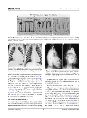
Figure 14. Examples of Doppler studies before (A), the next day following (B), and 8 months following (C) balloon pulmonary valvuloplasty (BPV)
demonstrating a reduction in Doppler peak instantaneous gradient from 92 mmHg (A) to 17 mmHg on the day after (B) and to 20 mmHg 8 months (C)
after BPV. Reproduced from Rao. 52
A B A B
Figure 15. Chest roentgenograms obtained before (A) and at intermediate-
term follow-up (B) after balloon pulmonary valvuloplasty demonstrate a Figure 16. Cineangiograms of the right ventricle (RV) captured before
decrease in the diameter of the cardiac silhouette. Adopted from Rao. 19 (A) and at intermediate-term follow-up (B) after balloon pulmonary
valvuloplasty demonstrate the total resolution of tricuspid valve
the pulmonary valve gradients decreased (98 ± 45 mmHg vs. regurgitation. The pulmonary artery (PA) and right atrium (RA) are
46 ± 33 mmHg; P < 0.05) following the first BPV (Figure 21). labeled. Adopted from Rao. 19
These patients were restudied 11 months (on average) later;
the pulmonary valve gradients increased (89 ± 40 mmHg; to be dilated with one balloon. When the double-balloon
P < 0.05) and were similar (P > 0.1) to pre-BPV values. method is utilized, the effective balloon diameter may be
Subsequent repeat BPV resulted in a significant reduction determined by Equation I:
(P < 0.01) of pulmonary valve gradients from 89 ± 40 mmHg Effective balloon diameter = 0.82 (D1 + D2) (I)
to 38 ± 20 mmHg (P < 0.01). Doppler studies conducted 2 Where D1 and D2 represent balloon diameters used
– 6½ years following repeat BPV showed excellent findings during BPV. The formula to compute the effective diameter
with residual Doppler-derived gradients of 24 ± 13 mmHg of both balloons together was developed by Rao and later
55
(Figure 21). Based on these observations, it may be inferred simplified by Narang et al. Some cardiologists advocated
56
that repeating BPV is useful and valuable in relieving the use of the double-balloon technique, particularly in
recurrent narrowing of the pulmonary valve. 54 adult patients. Therefore, Rao and Fawzy investigated
57
58
whether the double-balloon method is superior to using
2.7. Single- versus double-BPV
one balloon for BPV. As demonstrated in Figure 22, both
The double-balloon method (Figure 3) was employed for immediate and follow-up outcomes of the two-balloon and
BPV before the availability of balloons with large diameters one-balloon methods of BPV were excellent (P < 0.001)
in patients with an annulus of the pulmonary valve too big and similar (P > 0.1). The B/A ratios used for both
Volume 2 Issue 1 (2024) 6 https://doi.org/10.36922/bh.2406