
Page 20 - BH-2-1
P. 20
Brain & Heart Pictorial rendition pulmonary stenosis
A B
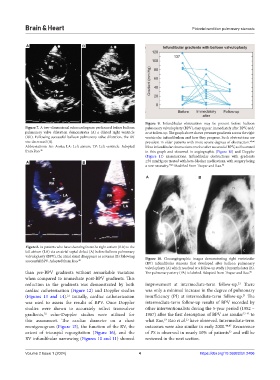
Figure 9. Infundibular obstruction may be present before balloon
Figure 7. A two-dimensional echocardiogram performed before balloon pulmonary valvuloplasty (BPV), may appear immediately after BPV, and/
pulmonary valve dilatation demonstrates (A) a dilated right ventricle or at follow-up. The graph above shows pressure gradients across the right
(RV). Following successful balloon pulmonary valve dilatation, the RV ventricular infundibulum and how they progress. Such obstructions are
size decreased (B). prevalent in older patients with more severe degrees of obstruction. 45,46
Abbreviations: Ao: Aorta; LA: Left atrium; LV: Left ventricle. Adopted Most infundibular obstructions resolve after successful BPV, as illustrated
from Rao. 49 in this graph and observed in angiographic (Figure 10) and Doppler
(Figure 11) examinations. Infundibular obstructions with gradients
A B ≥50 mmHg are treated with beta-blocker medications, with surgery being
a rare necessity. 45,46 Modified from Thapar and Rao. 50
A B
Figure 8. In patients who have shunting from the right atrium (RA) to the
left atrium (LA) via an atrial septal defect (A) before balloon pulmonary
valvuloplasty (BPV), the atrial shunt disappears or reverses (B) following Figure 10. Cineangiographic images demonstrating right ventricular
successful BPV. Adopted from Rao. 48
(RV) infundibular stenosis that developed after balloon pulmonary
valvuloplasty (A) which resolved at a follow-up study 10 months later (B).
than pre-BPV gradients without remarkable variation The pulmonary artery (PA) is labeled. Adopted from Thapar and Rao. 50
when compared to immediate post-BPV gradients. This
reduction in the gradients was demonstrated by both improvement at intermediate-term follow-up. There
21
cardiac catheterization (Figure 12) and Doppler studies was only a minimal increase in the degree of pulmonary
(Figures 13 and 14). Initially, cardiac catheterization insufficiency (PI) at intermediate-term follow-up. The
21
21
was used to assess the results of BPV. Once Doppler intermediate-term follow-up results of BPV recorded by
studies were shown to accurately reflect transvalvar other interventionalists during the 5-year period (1982 –
gradients, echo-Doppler studies were utilized for 1987) after the first description of BPV are similar 22-42 to
52
19
21
this assessment. The cardiac diameter on a chest what Rao, Rao et al. have observed. Intermediate-term
roentgenogram (Figure 15), the function of the RV, the outcomes were also similar in early 2000. 44,47 Recurrence
extent of tricuspid regurgitation (Figure 16), and the of PS is observed in nearly 10% of patients and will be
53
RV infundibular narrowing (Figures 10 and 11) showed reviewed in the next section.
Volume 2 Issue 1 (2024) 4 https://doi.org/10.36922/bh.2406