
Page 57 - GPD-2-4
P. 57
Gene & Protein in Disease K fragment for resistance gene hunting
(Thermo Scientific , USA) was used. Briefly, 10 µl of DNA, and final elongation at 72°C for 5 min in a reaction volume
™
2 µl of 10× buffer, 1 µl of T4 DNA Ligase (5 Weiss U/µl), and of 50 µl. PCR was executed in a T100 thermal cycler (Bio-
7 µl of water were mixed. The mixtures were incubated at rad laboratories, Inc, USA). The sequences of primers used
™
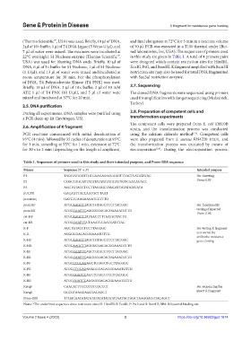
22°C overnight. S1 Nuclease enzyme (Thermo Scientific , in this study are given in Table 1. A total of 4 primers pairs
USA) was used for blunting DNA ends. Briefly, 10 µl of were designed which contain restriction sites for HindIII,
DNA, 6 µl of 5× buffer for S1 Nuclease, 1 µl of S1 Nuclease EcoRI, PstI, and BamHI. K fragment amplified with BamHI
(1 U/µl), and 13 µl of water were mixed and incubated at restriction site may also be used for total DNA fragmented
room temperature for 30 min. For the phosphorylation with Sau3aI restriction enzyme.
of DNA, T4 Polynucleotide Kinase (T4 PNK) was used.
Briefly, 10 µl of DNA, 2 µl of 10× buffer, 2 µl of 10 mM 2.7. Sequencing
ATP, 1 µl of T4 PNK (10 U/µl), and 5 µl of water were The cloned DNA fragments were sequenced using primers
mixed and incubated at 37°C for 20 min. used for amplification with Sanger sequencing (Medsantek,
Turkey).
2.5. DNA purification
During all experiments, DNA samples were purified using 2.8. Preparation of competent cells and
a PCR clean-up kit (Invitrogen, US). transformation experiments
The competent cells were prepared from E. coli DH10B
2.6. Amplification of K fragment strain, and the transformation process was conducted
PCR reactions commenced with initial denaturation at using the calcium chloride method . Competent cells
[14]
95°C (4 min), followed by 35 cycles of denaturation at 95°C were also prepared from S. aureus RN4220 strain, and
for 1 min, annealing at 55°C for 1 min, extension at 72°C the transformation process was executed by means of
for 30 s to 2 min (depending on the length of amplicon), electroporation . During the electroporation process,
[14]
Table 1. Sequences of primers used in this study and their intended purpose, and Prom-RBS sequence
Primer Sequence (5’ → 3’) Intended purpose
P1 TATATATATATTGTCAACAGACCAAGTTTACTCATATATAC For inserting
P2 CGGCTAGCATTATATATATATATATATTGTCAACAGACC Prom-RBS
P3 AGCTGTACCTCCTTACGGCTAGCATTATATATATATA
pUCPR GACAGTTACCAATGCTTAAT
prmtrSeq GATCTCAAGAAGATCCTTTG
promHF ATGCAAGCTTAGCTGTACCTCCTTACGGC For functionality
promER ATGCGAATTCAGGGCGACACGGAAATGTTG testing of inserted
Prom-RBS
cat-HF ATGCAAGCTTATGACTTTTAATATTATTG
cat-ER ATGCGAATTCCTAAATCCAATCATCTAC
K-F AGCTGTACCTCCTTACGGC For testing K fragment
K-R AGGGCGACACGGAAATGTTG as a vector for
antibiotic resistance
K-HF ATGCAAGCTTAGCTGTACCTCCTTACGGC gene cloning
K-HR ATGCAAGCTTAGGGCGACACGGAAATGTTG
K-EF ATGCGAATTCAGCTGTACCTCCTTACGGC
K-ER ATGCGAATTCAGGGCGACACGGAAATGTTG
K-PF ATGCCTGCAGAGCTGTACCTCCTTACGGC
K-PR ATGCCTGCAGAGGGCGACACGGAAATGTTG
K-BF ATGCGGATCCAGCTGTACCTCCTTACGGC
K-BR ATGCGGATCCAGGGCGACACGGAAATGTTG
KseqF CAACATTTCCGTGTCGCCCT For sequencing the
KseqR GCCGTAAGGAGGTACAGCT insert K fragment
Prom-RBS TTGACAATATATATATATATATATATAATGCTAGCTAAGGAGGTACAGCT
Notes: *The underlined sequences show restriction sites; H: HindIII; E: EcoRI; P: PstI; and B: BamHI; RBS: Ribosomal binding site.
Volume 2 Issue 4 (2023) 3 https://doi.org/10.36922/gpd.1674