
Page 60 - GPD-2-4
P. 60
Gene & Protein in Disease K fragment for resistance gene hunting
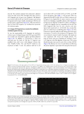
selected. The resistant colonies were subjected to plasmid and K-HR, K-EF and K-ER, K-PF and K-PR, and K-BF
extraction and colony PCR, through which the presence and K-BR primer pairs (Table 1). The genomic DNA was
of K fragment and cat gene was confirmed. The plasmid digested by HindIII, EcoRI, PstI, and Sau3aI enzymes and
was cleaved with EcoRI and HindIII enzymes, generating K fragment was digested by the same enzymes, except for
K fragment and cat gene bands, measuring at 1861 bp and Sau3aI, for obtaining compatible cohesive ends (Figure 6B).
626 bp in sizes, respectively (Figure 5C). This result showed K fragment was digested by BamHI to obtain overhangs at
that the Prom-RBS sequence can facilitate the expression 5’ ends of K fragment that are compatible with Sau3aI. The
of a resistance gene. DNA fragments that have the same cohesive ends were
ligated and transferred to E. coli DH10B. The colonies that
3.3. Usage of K fragment as a vector for antibiotic grew on erythromycin-containing plate (Figure 7A) were
resistance gene cloning subjected to plasmid isolation and subsequent PCR-based
To test the employability of K fragment for antibiotic examination. To confirm the existence of K fragment, PCR
resistance gene cloning from genomic DNA, genomic DNA was performed using K-F and K-R primers. The amplicons
was isolated and subjected to agarose gel electrophoresis were sequenced with KseqF and KseqR primers. The
(Figure 6A). In addition to chromosome, a band was sizes of inserts that give erythromycin resistance cloned
observed near the level of 2500 bp. K fragment was in K fragment after the digestion of genomic DNA using
amplified from pKF vector with designed primers that HindIII enzyme were equal to each other at about 2400 bp
have HindIII, EcoRI, PstI, and BamHI restriction site (Figure 7B), whereas the sizes of inserts obtained following
sequences at their 5’ ends. The primers used are K-HF the partial digestion of genomic DNA using Sau3aI enzyme
A B C
Figure 5. Confirmation of the functionality of Prom-RBS on K fragment by cloning cat gene. (A) For the cloning of cat to K fragment according to the
desired orientation, EcoRI and HindIII restriction site sequences were added to the designed primers used for polymerase chain reaction (PCR). Using
primers containing the restriction site sequences, cat fragment could be cloned into K fragment in the desired orientation. (B) Confirmation of the
presence of K fragment and cat fragment in transformants obtained in colonies grown on an agar plate containing chloramphenicol. Lane 1: PCR amplicon
obtained from chloramphenicol-resistant transformants using cat-HF and cat-ER primers. Lane 2: PCR amplicon obtained from chloramphenicol-resistant
transformants using K-HF and K-ER primers. M line: Lambda-PstI marker. (C) K fragment Ω cat plasmid. Lane 1: Two bands representing the fragments
of cat (626 bp) and K fragment (2861 bp) generated after digestion of the plasmid with EcoRI and HindIII enzymes. Lane 2: Circular form of K fragment
Ω cat plasmid. M line: Lambda-PstI marker.
A B
Figure 6. Resistance gene cloning from genomic DNA into K fragment. (A) Genomic DNA of Staphylococcus aureus MRSA ADU2 strain. Lane 1: The
isolated total DNA and a band that was thought a plasmid with an approximate length of 2500 bp. M line: Lambda-PstI marker. (B) Electrophoresis results
of the genomic DNA of MRSA ADU2 strain and K fragment digested by HindIII, EcoRI, PstI, and Sau3AI (BamHI) enzymes. Lanes 1, 3, 5, and 7 represent
the electrophoresed products after the digestion of genomic DNA by HindIII, EcoRI, PstI, and Sau3aI, respectively. Lanes 2, 4, 6, and 8 represent the
electrophoresed products after the digestion of genomic DNA by HindIII, EcoRI, PstI, and BamHI, respectively.
Volume 2 Issue 4 (2023) 6 https://doi.org/10.36922/gpd.1674