
Page 33 - GTM-1-2
P. 33
Global Translational Medicine Proteomic analysis of heart in metabolic cardiomyopathy
chromatography system (Sciex Co.). The peptides were 2.7. Western blot
concentrated on a 1.0-cm precolumn (75-µm inner Total proteins from cells or tissue were lysed using RIPA
diameter, 360-µm outer diameter, C18 5 µm, Sciex). The buffer, and the protein concentration in the cell lysates was
peptides were eluted from the precolumn using a gradient assayed by a protein assay dye reagent concentrate (Bio-
from 100% phase A (0.1% fatty acid aqueous solution) to Rad, USA). Samples were separated by sodium dodecyl
45% phase B (0.1% fatty acid, 100% acetonitrile) in 75 min sulfate-polyacrylamide gel electrophoresis (SDS-PAGE)
at 300 nL/min directly onto an 15-cm analytical column and then transferred to polyvinylidene fluoride (PVDF)
(75-µm inner diameter, 360-µm outer diameter, ReproSil- membranes (pore size 0.45 µm). After being blocked with
Pur C18 3 µm, Sciex). The instrument was operated in a 5% bovine serum albumin for 1 h, the membranes were
data-dependent mode automatically. Three biological incubated with primary antibodies, including CPT1B
replicates were prepared for each sample using a described (1:1000), ACAA2 (1:1000), and tubulin (1:5000) at 4°C
parameters (2500V+). overnight. After being washed with TBST, the membranes
were incubated with secondary antibodies for 1 h at room
2.4. Data processing and assembly temperature. Finally, the protein bands were visualized
Raw files from LC-MS were searched using the Mascot by enhanced chemiluminescence kit (Santa Cruz, Texas,
search engine human database 3.78 for data processing. USA).
Parameters were adjusted for: (i) trypsin digestion,
with two maximum missed cleavage points permitted; 3. Results
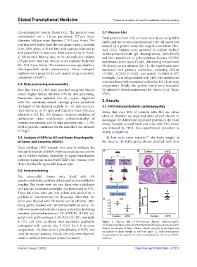
(ii) length of the digested peptide: 6 – 25; (iii) precursor 3.1. HFD-induced diabetic cardiomyopathy
mass tolerance of 10 ppm and fragment mass tolerance Given that over 80% of patients with MC are either
adjusted to 0.2 Da; (iv) dynamic variation oxidation of obese or diabetic, we employed diet-induced obesity to
methionine: static modification carbamidomethyl of investigate the differential expressed proteins in the heart
cysteine was selected; and (v) false discovery rate: < 1. The tissues between normal heart and mice with MC, which
level of peptide confidence for the data filter was adjusted was induced by HFD. The experimental procedure is
to “high.” shown in Figure 1A.
2.5. Analysis of DEPs by GO and Kyoto Encyclopedia In line with other reports, [15] the body weight of
of Genes and Genomes (KEGG) the mice in the HFD group almost doubled, and their
Gene ontology (GO) analysis was used to evaluate the
biological function of DEPs. Pathways analysis was carried A B
out to further evaluate metabolic or signal transduction
pathways using the online PANTHER tools (version 15.0)
(http://pantherdb.org/invalid Request.jsp).
2.6. Immunostaining
The myocardial tissues were fixed with 4%
paraformaldehyde, and then dehydrated and embedded in
paraffin. The hearts were cut into slices with a thickness
of 4 µm and incubated overnight in a thermostat at 37°C.
Then, the slices were put into xylene and alcohol (in a
gradient of concentration) for dewaxing. After that, the C
slices were blocked with 5% bovine serum albumin. After
being gently washed with phosphate-buffered saline, the
cells were incubated with the primary antibodies, including
carnitine palmitoyltransferase 1B (CPT1B) (1:100) and
acetyl-CoA acyltransferase 2 (ACAA2) (1:100), overnight
at 4°C, and then incubated with secondary antibodies Figure 1. High-fat diet (HFD)-induced diabetic cardiomyopathy.
conjugated with cyanine dye 3 (Cy3) for 1 h at room (A) Experimental procedure chart for this study. (B) Immunohistochemistry
temperature. 4’,6-diamidino-2-phenylindole (DAPI) was showed an increase of heart volume, cardiac myocyte hypertrophy and
an increase of heart weight in HFD-fed mice. (C) Echocardiographic
used for nuclear staining. Finally, the cells were observed measurement of the diastolic function in standard diet-fed mice or HFD-
under a confocal microscope (Leica, Germany). fed mice.
Volume 1 Issue 2 (2022) 3 https://doi.org/10.36922/gtm.v1i2.137