
Page 36 - GTM-1-2
P. 36
Global Translational Medicine Proteomic analysis of heart in metabolic cardiomyopathy
A
B
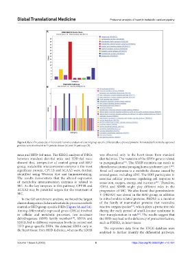
Figure 4. Kyoto Encyclopedia of Genes and Genomes analysis of control group-specific differentially expressed proteins. Annotated differentially expressed
proteins were distributed in 51 protein classes (A) and 33 pathways (B).
mice and HFD-fed mice. The KEGG analysis of DEGs was observed only in the heart tissue from standard
between standard diet-fed mice and HFD-fed mice diet-fed mice. The mutation of the SDHA gene is related
showed that, irrespective of control group and HFD to paraganglioma [19] . The SDHB mutation can result in
group, metabolite interconversion enzyme is the most pheochromocytoma/paraganglioma syndrome type 4 [20] .
significant protein. CPT1B and ACAA2 were, further, Renal cell carcinoma is a metabolic disease caused by
identified using Western blot and immunostaining. several genes, including SDH. The SDH participates in
The results demonstrate that the altered expression essential cellular processes regulating cell response to
of metabolite interconversion enzymes is related to sense iron, oxygen, energy, and nutrients [21] . Therefore,
MC. As the key enzymes in this pathway, CPT1B and SDHA and SDHB might play different roles in the
ACAA2 may be potential targets for the treatment of progression of MC. We also found that peroxiredoxin
MC. V (PRDX5) was absent in the HFD group, in addition
In the GO enrichment analysis, we found the largest to mitochondria-related proteins. PRDX5 is a member
cluster changes in cellular and metabolic processes in both of the family of mammalian proteins that neutralize
control or HFD group-specific DEPs (Figure 3A and 5A). reactive oxygen species [22] , which plays a protective role
Among differentially expressed genes (DEGs) involved during the early period of small-for-size syndrome in
in cellular and metabolic processes, two succinate liver transplantation in rats [23] . The results suggest that
dehydrogenase (SDH) family members [18] , SDHA and the HFD may lead to the deficiency of protective factors,
SDHB, led to different expression levels in control and such as PRDX5, in heart tissue.
HFD group-specific DEPs. We detected SDHA only in
the heart tissue from HFD-fed mice, whereas the SDHB The expression data from the KEGG database were
analyzed to further identify the differential pathways.
Volume 1 Issue 2 (2022) 6 https://doi.org/10.36922/gtm.v1i2.137