
Page 34 - GTM-2-3
P. 34
Global Translational Medicine Deep learning by NMR-biochemical
A B
C D
E F
G H
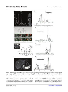
Figure 1. Representative nuclear magnetic resonance (NMR) peaks are used in magnetic resonance spectroscopic (MRS) imaging methods and biochemical
tests by assessing metabolite concentrations in tissue and serum to interpret the disease burden in normal brain, breast, myocardium, prostate, and liver.
(A-D) are NMR of the brain, heart, muscle, and liver, respectively, and (E-H) are the MRS results of the respective organs. Images are reproduced from a
dissertation submitted at IIT Delhi in 1995 .
[1]
different locations in human tissues for physiological and robust automated MRS imaging (MRSI) segmentation
functional metabolic screening with parallel development and registration processing have been routinely used for
in ultrahigh-resolution NMR imagers . Simultaneously, extracting focal lesion features (size, chemical composition,
[2]
Volume 2 Issue 3 (2023) 2 https://doi.org/10.36922/gtm.337