
Page 45 - IJB-1-1
P. 45
Makoto Nakamura, Tanveer A. Mir, Kenichi Arai, et al.
cation in a single molecule, are known as excellent and 1% penicillin and streptomycin (P/S), as well as
bio-functional materials that prevent both cell D-glucose and essential saline. SMCs were seeded
5
adhesion and protein adsorption [17,18] , and are also onto stripe patterned discs at a concentration of 7×10
useful for bio-patterning [19,20] . In this work, zwitte- cells per one well in 24-well plates and cultured in
rionic polymers were prepared using 1-carboxy-N,N- Dulbecco’s Modified Eagle Medium (DMEM, Gibco,
dimethyl-N-(2’-methacryloyloxyethyl)methanaminium USA) supplemented with 10% FBS and 1% P/S. The
inner salt also known as carboxymethyl betaine (CMB) cells were cultured at 37℃ with 5% CO 2 in an incu-
was kindly donated from Osaka Organic Chemical bator and the medium was changed every two days.
Industry, Kashiwara, Japan, and n-butylmethacrylate
(BMA) obtained from Wako, Osaka, Japan, at the rate 2.3 Ethics Statement
of (CMB/BMA = 29/71; MW = 193 kDa and MW/Mn All animal experiments were performed according to a
=1.92; 1 wt% ethanol solution), and utilized as a protocol approved by the Committee on Ethics in
surface patterning material. 1% CMB ethanol solution Animal Experiments of the University of Toyama,
was used to print stripe patterns onto plastic culture Japan.
discs with a custom-made inkjet 3D Bioprinter [21–24]
equipped with an inkjet head (IJHB-1000, MICRO- 2.4 Patterning of Cells and Transfer Printing
JET Corporation, Nagano, Japan). The width of each After being seeded and cultured on stripe patterned
printed line was 200 µm with an interval width be- coated discs, the cells attached only to the CMB/BMA
tween lines of 500 µm. After printing, the patterned non-coated areas, with linear patterned CMs, MBs,
discs were dried and sterilized by UV irradiation be- and SMCs were obtained. Next, we performed transfer
fore being used in the experiments. printing on day 9 for CMs, day 7 for MBs, and day 1
2.2 Preparation of Muscle Cells for SMCs after seedings. In this procedure, culture
discs with patterned cells were taken from the wells,
Primary rat neonatal cardiomyocytes (CMs) and turned upside down and placed on a Matrigel substrate
myoblasts (MBs) were obtained from 1 day old neo- (BD Biosciences, USA). After 12 hours of cultivation,
natal rats (Slc: Wistar). Smooth muscle cells (SMCs) the discs were removed and cells on the patterned
were purchased from the Japanese Collection of Re- discs which showed linear patterns were transferred to
search Bioresources (JCRB), Osaka, Japan. Matrigel substrates while maintaining their linear pat-
To prepare primary cells, cardiac and skeletal mus- terns, then cultured in succession and observed. These
cle tissues were excised from the upper and lower ex- procedures are shown in Figure 1. To prevent the
tremities and the heart of neonatal rats respectively transferred linear cell patterns from shrinking, we em-
and chopped into small pieces. The respective muscle ployed a custom-made silver wire (0.5 mm in diameter,
tissue pieces were treated with collagenase. Cell sus- Niraco, Tokyo, Japan) coated with type-I collagen
pensions of primary CMs and MBs were obtained. (Cellmatrix, Nitta Geratin Corp. Osaka, Japan) for
The obtained primary cells were seeded onto stripe anchoring the ends of the patterns (Figure 2). In brief,
6
patterned discs at a concentration of 1×10 cells per the silver wires were first treated with type-I collagen,
5
one well in 12-well plates or 7×10 cells per one well then inserted on the border areas of cell patterns on
in 24-well plates, then cultured in Medium 199 (M199) Matrigel using manual autoclaved forceps. It was ex-
supplemented with 10% fetal bovine serum (FBS), pected that cells will cover the type-I collagen coated
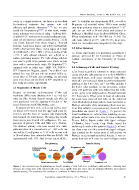
Figure 1. The procedures used for patterning and transfer printing of cells. Cells were seeded onto patterned discs. Following their
adhesion, the culture discs with patterned cells were turned upside down and placed onto the Matrigel substrates, then removed
(transfer printing). The transferred cells continued to be cultured for further observations.
International Journal of Bioprinting (2015)–Volume 1, Issue 1 41