
Page 47 - IJB-1-1
P. 47
Makoto Nakamura, Tanveer A. Mir, Kenichi Arai, et al.
was unchanged in most cases. In addition, spontane-
ous contractions by the CMs were also observed, al-
though those were limited to only partial portions of
the fiber-like structures. However, in the case of CMs
and MBs, it was observed that some of the cells had
spread to the Matrigel substrate.
The use of silver wire as an anchor was signifi-
cantly effective in maintaining the fiber-like structures.
The design and representative photograph are shown
in Figure 2. Without use of an anchor, fiber-like struc-
tures produced with transferred cells became wrinkled
over time, and finally formed tangled or aggregated
groups of cells after 24 hours (Figure 6(A-1), 6(A-2)),
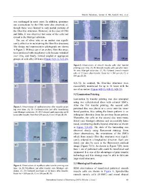
Figure 5. Observations of smooth muscle cells after transfer
printing over time. (A, B) Smooth muscle cells just after trans-
fer onto Matrigel substrates. (C, D) Cultured smooth muscle
cells at 12 hours after transfer. Scale bar = 200 µm (A, C) or
100 µm (B, D).
6(A-3)). In contrast, the fiber-like structures were
successfully maintained for up to 36 hours with the
use of an anchor (Figure 6(B-1), 6(B-2), 6(B-3)).
3.2 Lamination Printing
Lamination by transfer printing was also attempted
using two cell-patterned discs with cultured SMCs.
After the first transfer printing, the second cell-
Figure 3. Observations of cardiomyocytes after transfer print-
ing over time. (A, B) Cardiomyocytes just after transferring patterned disc was placed as a cover onto the trans-
onto Matrigel substrates. (C, D) Cultured cardiomyocytes at 12 ferred position, thus setting the linear patterns in an
hours after transfer. Scale bar=200 µm (A, C) or 100 µm (B, D). orthogonal direction from the previous linear pattern.
Thereafter, the cells on the second disc were trans-
ferred onto Matrigel substrate and successfully lami-
nated, constructing double layered structures as shown
in Figure 7(A–D). The state of the laminating was
observed clearly using fluorescent staining. From
closer observations, the orientations of the SMCs
which form muscle fiber-like structures were signifi-
cantly ordered in a longitudinal direction. Such ten-
dency can also be seen in the fluorescent confocal
image (Figure 7(C)). As shown in Figure 7(D), more
than 1 cm of patterned cells could be transferred and
laminated. It is one of the advantages of print transfer
technology and this strategy may be able to fabricate
large sized structures.
3.3 Histological Evaluation
Figure 4. Observations of myoblasts after transfer printing over
time. (A, B) Myoblasts just after transfer onto Matrigel sub- SEM observations of transferred patterned smooth
strates. (C, D) Cultured myoblasts at 12 hours after transfer. muscle cells are shown in Figure 8. Spindle-like
Scale bar = 200 µm (A, C) or 100 µm (B, D). smooth muscle cells (S-SMC) and round shaped
International Journal of Bioprinting (2015)–Volume 1, Issue 1 43