
Page 46 - IJB-1-1
P. 46
Bioprinting with pre-cultured cellular constructs towards tissue engineering of hierarchical tissues
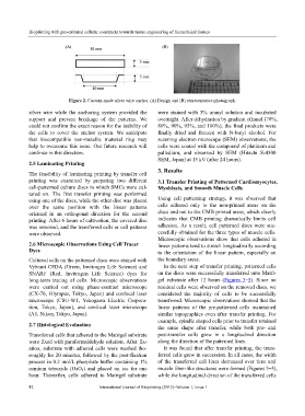
Figure 2. Custom-made silver wire anchor. (A) Design and (B) representative photograph.
silver wire while the anchoring system provided the were stained with 3% uranyl solution and incubated
support and prevent breakage of the patterns. We overnight. After dehydration by gradient ethanol (70%,
could not confirm the exact reason for the inability of 80%, 90%, 95%, and 100%), the final products were
the cells to cover the anchor system. We anticipate finally dried and freezed with N-butyl alcohol. For
that biocompatible non-metallic material ring may scanning electron microcope (SEM) observations, the
help to overcome this issue. Our future research will cells were coated with the compound of platinum and
continue in this direction. palladium, and observed by SEM (Hitachi S-4500
SEM, Japan) at 15 kV (after 24 hours).
2.5 Laminating Printing
3. Results
The feasibility of laminating printing by transfer cell
printing was examined by preparing two different 3.1 Transfer Printing of Patterned Cardiomyocytes,
cell-patterned culture discs in which SMCs were cul- Myoblasts, and Smooth Muscle Cells
tured on. The first transfer printing was performed
using one of the discs, while the other disc was placed Using cell patterning strategy, it was observed that
over the same position with the linear patterns cells adhered only to the non-printed areas on the
oriented in an orthogonal direction for the second discs and not to the CMB printed areas, which clearly
printing. After 6 hours of cultivation, the covered disc indicates that CMB printing dramatically limits cell
was removed, and the transferred cells or cell patterns adhesion. As a result, cell patterned discs were suc-
were observed. cessfully obtained for the three types of muscle cells.
Microscopic observations show that cells adhered in
2.6 Microscopic Observations Using Cell Tracer linear patterns tend to stretch longitudinally according
Dyes to the orientation of the linear pattern, especially on
Cultured cells on the patterned discs were stained with the boundary areas.
Vybrant CFDA (Green, Invitrogen Life Science) and In the next step of transfer printing, patterned cells
SNARF (Red, Invitrogen Life Science) dyes for on the discs were successfully transferred onto Matri-
long-term tracing of cells. Microscopic observations gel substrate after 12 hours (Figures 3–5). Since no
were carried out using phase-contrast microscope residual cells were observed on the removed discs, we
(CX-70, Olympus, Tokyo, Japan) and confocal laser considered the majority of cells to be successfully
microscope (CSU-W1, Yokogawa Electric Corpora- transferred. Microscopic observations showed that the
tion, Tokyo, Japan), and confocal laser microscope linear patterns of the pre-patterned cells maintained
(A1, Nikon, Tokyo, Japan). similar topographies even after transfer printing. For
example, spindle shaped cells prior to transfer retained
2.7 Histological Evaluation
the same shape after transfer, while both pre- and
Transferred cells that adhered to the Matrigel substrate post-transfer cells grew in a longitudinal direction
were fixed with paraformaldehyde solution. After fix- along the direction of the patterned lines.
ation, substrate with adhered cells were washed tho- It was found that after transfer printing, the trans-
roughly for 20 minutes, followed by the post-fixation ferred cells grew in succession. In all cases, the width
process in 0.2 mol/L phosphate buffer containing 1% of the transferred cell lines decreased over time and
osmium tetroxide (OsO 4) and placed on ice for one muscle fiber-like structures were formed (Figures 3–5),
hour. Thereafter, cells adhered to Matrigel substrate while the longitudinal direction of the transferred cells
42 International Journal of Bioprinting (2015)–Volume 1, Issue 1