
Page 49 - IJB-1-1
P. 49
Makoto Nakamura, Tanveer A. Mir, Kenichi Arai, et al.
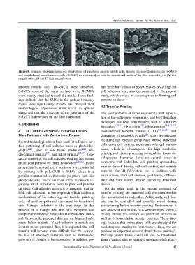
Figure 8. Scanning electron microscopic observations of transferred smooth muscle cells. Spindle like smooth muscle cells (S-SMC)
and round shaped smooth muscle cells (R-SMC) were observed on both the outside and inside of the fiber respectively at (A) low
magnification, (B and C) high magnification.
smooth muscle cells (R-SMCs) were observed. tent inhibitory effects of poly(CMB-co-BMA) against
S-SMCs covered the outer surface while R-SMCs cell adhesion were also demonstrated in the present
were mainly stratified toward the inside. These find- study, which should be advantageous to forming cell
ings indicate that the SMCs in the surface boundary patterns on discs.
region were significantly affected and changed their
morphological appearance from round to spindle 4.2 Transfer Printing
shape and that the direction of the long axis of the The great potential of tissue engineering with applica-
S-SMCs is dependent on the fiber’s direction. tion of bio-patterning, bioprinting, and bio-fabrication
techniques has been demonstrated, such as solid free
4. Discussion
formations [30,31] , 3D printing [32] , inkjet printing [2–4,21–24] ,
4.1 Cell Cultures on Surface Patterned Culture laser-induced forward transfer (LIFT) [10–12,33] , and
Discs Patterned with Zwitterionic Polymer dispensing of spheroids of cells . Many investigators
[5]
Several technologies have been used for effective sur- including our research group have printed individual
face patterning of cell cultures, such as photolitho- cells using cell printing techniques with cell suspen-
graphy [25] , laser or ion beam irradiation [26] , mi- sions, which is advantageous for high resolution
cro-contact printing [27] , and inkjet printing [28,29] . Gen- printing and shows promising potential for future de-
erally, control of the cell adhesive position has been a velopments. However, there are several issues to
major goal pursued by many researchers [26–29] . In the overcome with individual cell printing approaches,
present study, non-adhesive positions were controlled such as the cell density, cell–cell contact, and suitable
by printing with poly(CMB-co-BMA), which is a materials for 3D fabrication, etc. In addition, cells
popular commercial zwitterionic polymers just like must adhere, start cell division, proliferate, differen-
phosphobetaine. There has been active discussion re- tiate and form tissues, before becoming functional
garding which is better in order to print cell patterns tissues.
on discs: Cell adhesive materials or materials that in- On the other hand, in the present approach of
hibit cell adhesion. In our approach which utilizes a transfer printing, the patterned cells are transferred as
combination of bio-patterning and transfer printing, they are adhered to each other. Additionally, cell den-
cells cultured on patterned discs must be transferred sity can be controlled and possibly raised during
onto Matrigel substrate at the next stage. In this pre-culturing before transfer printing. Furthermore, it
process, it is thought that the cells recognize and was observed that muscle cells were arranged longitu-
compare the adhesive molecules in the attached mate- dinally during pre-cultures on patterned surfaces as
rials between the patterned disc and the Matrigel sub- well as 6 hours during transfer printing. These find-
strate before transfer. If cell-adhesive materials are ings indicate that pre-cultured cells are already diffe-
located on the patterned disc, it is expected that cell rentiating and starting to form tissues. Thus, we can
transfer will become more difficult. For this reason, propose an important concept about “tissue printing”.
the use of inhibitory materials such as zwitterionic Partially grown tissue constructs can be transferred
polymers is thought to be reasonable. In addition, po- from a culture disc to Matrigel substrate while main-
International Journal of Bioprinting (2015)–Volume 1, Issue 1 45