
Page 64 - IJB-1-1
P. 64
A novel 3D printing method for cell alignment and differentiation
cence microscopy. minutes, then permeabilized with 0.25% triton X 100
for 10 minutes and washed with PBS, then blocked
2.5 Evaluation of Patterned Surface for Cell Alignment with 0.5% bovine serum albumin (BSA) for 30
5
The cells were seeded with a density of 3 × 10 minutes, all performed at 4°C. Cells were then incu-
2
cells/cm and left to grow for 2 days. For an evaluation bated with primary antibodies for 30 minutes, fol-
of cell orientation, the samples are stained with fluo- lowed by the FITC labeled secondary antibody for 30
rescein diacetate (FDA) and 4',6-Diamidino-2-PheNy- minutes. Cells were finally re-suspended in PBS and
lindole, Dihydrochloride (DAPI) nuclear stain. The analyzed for marker expression with the Millipore
cells are then observed under a fluorescence micro- GUAVA easycyte HT flow cytometer.
scope (Olympus IX71, Japan).
3. Results
2.6 Cardiac Expression Study for Aligned Cells on
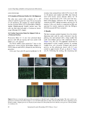
a Patterned Surface The back pressure-assisted dispenser from the robotic
dispensing unit could be easily substituted from the
Polystyrene films (2 × 2 cm) were patterned linear printing arm and replaced with a sharp needle that
grooves with 100 µm spacing and were seeded with could etch distinct grooves into polystyrene sheets
5
2
hMSC at 3 × 10 cells/cm . (Figure 1(A), (B), (C)). The apparatus could then be
For FACS, hMSCs were seeded for 7 days on the programmed to print complex patterns by creating
polystyrene surfaces before intracellular staining for straight lines, and sinusoidal S-shaped and circular
CD29 (Abcam) and GATA4 (Abcam) by the following patterns on the polystyrene sheets with X-Y axis
procedure: control to approximately 50 µm as shown in Figure
Cells were fixed with 4% paraformaldehyde for 30 2(A–F). Following the etching, the dispensing unit
Figure 2. Patterns etched into polystyrene films presented at x4 (A, B, C) and x10 (D, E, F) magnification. The robot could be pro-
grammed to etch linear (A and D), S-shaped or waveform (B and E) and concentric circles (C and F). The robotic dispensing system
etched a pattern of aligned grooves into the polystyrene surface (G), then printed the bioink directly to the grooves following identic-
al coordinates (H).
60 International Journal of Bioprinting (2015)–Volume 1, Issue 1