
Page 63 - IJB-1-1
P. 63
Ramya Bhuthalingam, Pei Qi Lim, Scott A. Irvine, et al.
Dulbecco's modified eagle's medium (DMEM), fet- ing robot (SANEI-TECH, Japan) and manufacturer
al bovine serum (FBS), and penicillin streptomycin supplied software. Modifications were made to the
solution were purchased from Invitrogen. robot to allow for ablation patterning. Specifically, the
dispensing head was replaced by a sharpened stylus in
2.2 Robotic Dispensing System order to cut the substrate surface (Figure 1).
The Janome 2300N pressure-controlled robotic dis- Since both the polystyrene tissue culture plates and
pensing system consists of a computer control, robotic polystyrene films were used, all surfaces were
XYZ table, and a pressure-driven syringe mechanism plasma-treated and coated with fetal bovine serum to
(Figure 1). Programming was done with the help of a produce a standardized surface treatment between ex-
specialized JR-C points software (manufacturer sup- periments. Plasma treatment, using a Femto Science
plied) to define the specifications for both dispensing system, preceded using conditions of 150 W, 30 sscm
and etching. Original programs (data points for gene- of oxygen for 10 minutes in a plasma system (Femto
rating various shapes) were created in compatible Science). FBS was then coated on the surface and in-
programming languages (such as DXF, Gerber data, cubated for 2 hours before washing thrice in PBS.
etc. in ASCII format) by using point job commands to Different patterns are generated by entering coor-
create the lines. Hence, the software controlled the dinate points into the JR-C points software. Patterns in
X-Y-Z geometry and the deposition rate. Bioink- a variety of forms were obtained for this study, e.g.
Printing was performed using 0.05 MPa back pressure, linear, S-shaped, and circle. Linear patterns with the
5 mm/sec writing speed from a 25 mL syringe and a spacing of 50, 100, 250, 500, and 1000 μm were
30 gauge needle (inner diameter 250 micron). produced. The depth of each groove can also be varied
easily using the Z-height of the system. In this test,
grooves set at 40, 80, and 170 μm were cut into the
surface of 1 mm thick polystyrene (PS) sheets.
In order to view the cross-section of the patterned
surface (especially for characterizing the depth of the
sample), the patterned sample was first scored along
the back of the samples to introduce a crack. Force
was then applied to produce a controlled stress frac-
ture across the sample. The exposed cross-section can
then be analyzed accurately. In order to visualize the
depth of the etching on the sample, an optical micro-
scope was used (Olympus IX71, Japan). For quantitative
measurement of the features (e.g. such as in measuring
the depth of the grooves), ImageJ software was used.
2.4 Preparation of Cell-containing Gelatin Bioink
Bioink solutions were prepared by dissolving gelatin
at 2% in DMEM. The solutions were heated and
stirred at 60°C for 2 hours to aid solubilization. After
cooling, the fibroblasts and the MSCs were suspended
6
−1
at concentration of 5 × 10 cells mL within the bio-
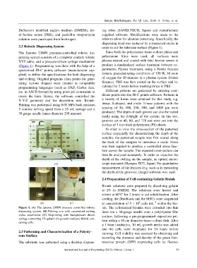
Figure 1. (A) The Janome 2300N pressure controlled robotic ink. The cellularized bioinks were extruded into thin
dispensing system. (B) Printing arm with customized etching lines via a 30-gauge needle onto a polystyrene film
stylus attachment. (C) Bioprinting with backpressure driven surface, following a pre-programmed deposition pat-
syringe containing 2% gelatin (in growth medium) bioink con-
taining cells. tern within a 10 cm diameter tissue culture dish. After
a 1 hour incubation, 10 mL growth media was added
and the cells were incubated for 24 hours before
2.3 Patterning and Characterization of a Polysty- viewing. Cell viability was assessed by observing and
rene Surface
recording the presence and density of the green fluo-
The substrate was patterned using a desktop dispens- rescence protein (GFP) expressing cells by fluores-
International Journal of Bioprinting (2015)–Volume 1, Issue 1 59