
Page 67 - IJB-1-1
P. 67
Ramya Bhuthalingam, Pei Qi Lim, Scott A. Irvine, et al.
incubated at 37°C. The stem cells sense the features onto these grooves. The printed cells can sense the
and then become stretched and aligned in the direction grooves and respond by elongating and aligning with
of the feature. Figure 7(A) demonstrates the high den- them. There is the potential of combining both proce-
sity of cells printed directly on to the etched grooves, sses in the same printing head to produce cell aligning
as can be viewed by the DAPI stained nuclei. The grooves and simultaneously seed the cells directly on
printed MSCs were then observed to adopt the the features as they are generated (Figure 8).
stretched morphology, aligning along the direction of
the etched groove (Figure 7(B)). When the MSCs are
deposited in the culture media rather than the bioink,
the cells do not form distinct traces; instead the cells
adhere both in between and within the grooves,
leading to a total confluence of the surface. The cells
seeded in this manner still become elongated and align
in the direction of the etched features, however, lack
the distinct printed trace along the grooves
(Figure 7(C)). Figure 7(D) displays non-elongated and
randomly aligned stem cells seeded on an unpatterned
surface as a control.
Figure 8. Proposed dual etching and bioprinting of a hard po-
lymer surface using a similar automated robotic dispenser.
The method described here presents a straightfor-
ward and time-efficient method to produce cell align-
ing features and to also cellularize with relative preci-
sion so that etching and bioprinting can both be per-
formed under an hour. Other methods of producing
aligning channels and grooves include deep reactive
ion etching [26] , electron beam lithography [12] , direct
laser writing [27] , femtosecond laser [22] , photolithogra-
phy [28] , plasma dry etching [29] etc. (as reviewed by Li
[7]
and colleagues ). These tend to be more time-consu-
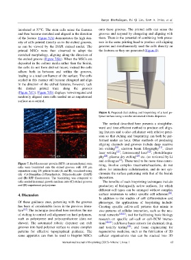
Figure 7. Red fluorescent protein (RFP) rat mesenchymal stem ming, involve complex treatments/reactions, do not
cells were bioprinted onto the etched grooves with 100 µm
separation using 2% gelatin bioink (A and B), visualized using allow for immediate cellularization, and do not syn-
(A) 4',6-Diamidino-2-Phenylindole, Dihydrochloride (DAPI) chronize the surface patterning with that of the bioink
and (B) RFP fluorescence. The bioprinting was compared to deposition.
cells seeded in normal growth medium onto (C) etched grooves The benefits of such bioprinting techniques include
and (D) unpatterned polystyrene. production of biologically active surfaces, for which
different cell types can be arranged without complex
4. Discussion surface treatments to select specific cell adhesion [30] .
In addition to the studies of cell differentiation and
Of these guidance cues, patterning with the grooves phenotype, the applications of bioprinting include:
has been of considerable focus in the previous litera- Creating specific cell-to-cell patterns that mimic in
ture [6,7] .The technique described here involves the use vivo patterns of cellular interaction, such as the neu-
of etching to control cell alignment on hard polymers, ronal networks [30,31] ; tool for facilitating basic biology
such as polystyrene and polycaprolactone (data not research on specific cell–cell or cell–ECM interac-
shown). The automated robotic dispenser can etch tions [30,32] ; cell/tissue bases sensors for chemical, drug,
grooves into hard polymer surface to create complex and toxicity testing [33] ; and tissue engineering for
patterns for effective topographical guidance. The regenerative medicine, such as the fabrication of 2D
same apparatus can then be used to bioprint MSCs cellular organizations that can be stacked into 3D
International Journal of Bioprinting (2015)–Volume 1, Issue 1 63