
Page 82 - IJB-1-1
P. 82
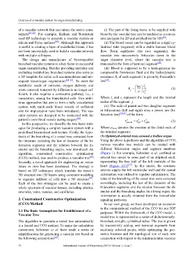
Creation of a vascular system for organ manufacturing
of a vascular network that can mimic the native coun- (i) The part of the living tissue to be supplied with
terparts [21,22] . For example, Kaihara and Borenstein blood by the vascular tree can be modeled as a convex
used RP technology to generate a vascular system on area (polygon for 2D and polyhedron for 3D) [27] .
silicon and Pyrex surfaces [23] . Although this approach (ii) The blood vessel can be regarded as a rigid cy-
is useful in creating a layer of endothelial tissue, it has lindrical tube (segment) with a stable laminar blood
not been successfully used to build a vascular network flow. From capillaries (the root segments), the
with multiple cell types. vascular tree successively bifurcates down to the
The design and manufacture of biocompatible larger diameter level, where the vascular tree is
branched vascular systems is a key factor in successful truncated in the form of terminal segment [28] .
organ manufacturing. Besides providing nutrients and (iii) Blood is supposed to be a homogeneous in-
excluding metabolites, branched systems also serve as compressible Newtonian fluid and the hydrodynamic
a 3D template for initial cell accommodation and sub- resistance R j of each segment j is given by Poiseuille’s
sequent tissue/organ regeneration [21–23] . To meet the law [29] :
metabolic needs of nutrients, oxygen delivery, and 8η l j
waste removal, transport by diffusion is no longer suf- R = π r 4 (1)
j
ficient; it also requires a convective pathway, i.e., a j
vasculature, among the transplanted cells. Two ambi- Where l j and r j represent the length and the internal
tious approaches that aim to form a fully vascularised radius of the segment j.
system with multi-scale blood vessels of sufficient (iv) The radii of parent and two daughter segments
size for implantation have been introduced. The vas- at bifurcations (left and right) obey a power law (bi-
cular systems are designed to be connected with the furcation law) [30,31] of the form:
patient’s own blood vessels during surgery [24] . r γ = r + r γ (2)
γ
In this perspective, we describe the two basic strat- parent left right
egies for producing a complex vascular system with a Where p term denotes the pressure at the distal ends of
predefined hierarchical architecture. Firstly, the hypo- the terminal segments.
thesis of the branching of a vessel tree, extracted from (1) Optimized arterial trees around a hollow organ
the literature including the law of diameter of the bi- Using the above hypotheses and the algorithm of CCO,
furcation segments and the relation between the di- various vascular tree models can be created with
ameter and the branching angles, was introduced. An different bifurcation angles and segment numbers
algorithm, constrained constructive optimization (Figure 1). For example, Schreiner et al. confined an
(CCO) method, was used to produce a vascular tree [25] . arterial tree model to some part of an elliptical shell,
Secondly, a novel approach for engineering as vascu- representing the free wall of the left ventricle of the
lature in vitro has been developed. The strategy is heart (Figure 1(C)) [32] . In this model, the coronary
based on RP techniques which translate the tissue’s arteries supply the left ventricular wall and the spatial
3D structure into 2D layers using computer modeling information was utilized to regulate optimization. The
to organize millions of cells into a 3D structure [26] . rules of the branching of the vessel tree were extracted
Each of the two strategies can be used to create a accordingly, including the law of the diameter of the
whole spectrum of vascular tissues, including arteries, bifurcation segments and the relation between the di-
arterioles, veins, venules, and capillaries. ameter and the branching angles. In a living organ, the
information is usually obtained from the intercellular
2. Constrained Constructive Optimization signaling pathways.
(CCO) Method In our own group, we have developed an extension
to the computational method of the CCO for our 3DP
2.1 The Basic Assumptions for Establishment of a purposes. Within the framework of the CCO model, a
Vascular Tree
model tree is represented as a series of dichotomously-
The algorithm to generate a vessel tree automatically branched, straight, cylindrical tubes. The tree is grown
is a fractal and CCO method. To make the calculation by successively adding new terminal segments from
convenient, Schreiner et al. have made a series of randomly selected points, while optimizing the geo-
simplifications for generating a vascular tree based on metric location and the topological site of each new
the following assumptions [27] . connection with respect to the minimum intra-vascular
78 International Journal of Bioprinting (2015)–Volume 1, Issue 1