
Page 156 - IJB-10-1
P. 156
International Journal of Bioprinting 3D-printed micro-perfused culture device
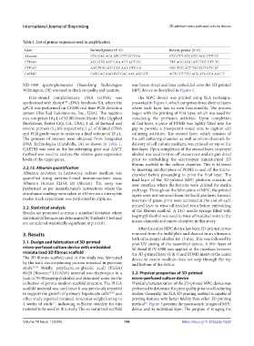
Table 1. List of primer sequences used in amplification
Gene Forward primer (5’-3’) Reverse primer (3’-5’)
Albumin CTG CAC AGA ATC CTT GGT GA CTC CTT ATC GTC AGC CTT GC
CYP3A4 ACC GTG ACC CAA AGT ACT GG TTC AGG GGG ATC TGT GTT TC
CYP3A7 AAG TGG ACC CAG AAA CTG CA GGC TCC ACT TAC GGT CTC AT
GAPDH CAT GAG AAG TAT GAC AAC AGC CT AGT CCT TCC ACG ATA CCA AAG T
ND-1000 spectrophotometer (NanoDrop Technologies was freeze-dried and later embedded onto the 3D-printed
Wilmington, DE) was used to check its quality and quantity. MPC device as described in Figure 1.
First-strand complementary DNA (cDNA) was The MPC device was printed using SLA technique,
synthesized with iScript cDNA Synthesis Kit, where the presented in Figure 1, which comprises three distinct layers
TM
qPCR was performed on CFX96 real-time PCR detection where each layer has its own functionality. The process
system (Bio-Rad Laboratories, Inc., USA). The reaction began with the printing of first layer, which was used for
mix comprises 10 µL of SYBR Green Master Mix (Applied visualizing the perfusion activities. Upon completion
Biosystems, Foster City, CA, USA), 1 µL of forward and of first layer, a piece of PDMS was tightly fitted into the
reverse primers (5 µM) respectively, 1 µL of diluted cDNA gap to provide a transparent visual area to capture cell
and PCR grade water to make up a final volume of 20 µL. culturing activities. The second layer, which consists of
The primers of interest were obtained from Integrated the cell culturing chamber as well as micro-channels for
DNA Technologies (Coralville, IA) as shown in Table 1. delivery of cell culture medium, was printed on top of the
GAPDH was used as the housekeeping gene and ∆∆CT first layer. Upon completion of the second layer, isopropyl
method was used to analyze the relative gene expression alcohol was used to rinse off excess resin and air gun dried
levels of the target genes. prior to embedding the electrospun miniaturized 3D
fibrous scaffold in the culture chamber. This is followed
2.2.13. Albumin quantification by inserting another piece of PDMS to seal off the micro-
Albumin secretion in hepatocyte culture medium was chamber before proceeding to print the final layer. The
quantified using enzyme-linked immunosorbent assay, final layer of the 3D-printed MPC platform consists of
Albumin Human ELISA kit (Abcam). The assay was user interface where the ferrules were printed for media
performed as per manufacturer’s instructions where the exchange. Throughout the fabrication of MPC, the printed
absorbance readings were taken at 450 nm on a microplate layers were not removed from the build platform. Instead,
reader. Each experiment was performed in triplicate. intervals of pause print were activated at the end of each
2.3. Statistical analysis printed layer to rinse off residual resin before embedding
Results are presented as mean ± standard deviation where of the fibrous scaffold. A 21G needle syringe filled with
statistical differences are determined by Student’s t-test and isopropyl alcohol was used to rinse off residual resin in the
are considered statistically significant at p ≤ 0.01. micro-channels and micro-chamber in this study.
After the entire MPC device has been 3D-printed, it was
3. Results removed from the build plate and cleaned in an ultrasonic
bath of isopropyl alcohol for 15 min. This was followed by
3.1. Design and fabrication of 3D-printed post-UV curing of the assembled device. A thin layer of
micro-perfused culture device with embedded Sil-Bond RTV-4500 was applied at the interface between
miniaturized 3D fibrous scaffold the 3D-printed layer (1 & 3) and PDMS insert of the cured
The 3D fibrous scaffold used in this study was fabricated device to ensure medium does not seep through the top
by the bath electrospinning process reported in previous and bottom of the device.
study. 45,46 Briefly, poly(lactic-co-glycolic acid) (PLGA)
80:20 (Resomer® LG 824S) material was electrospun in a 3.2. Physical properties of 3D-printed
bath of 70:30 isopropyl alcohol and deionized water for the micro-perfused culture device
collection of porous random scaffold structure. The PLGA Physical characterization of the 3D-printed MPC device was
scaffold material was used since it was previously reported performed to determine the print quality prior to cell culturing
to support the growth of primary hepatocyte cells 48-50 and studies. Generally, the SLA 3D printing method is capable of
other study reported minimal molecular weight lost up to printing features with better fidelity than other 3D printing
52
4 weeks of study , indicating sufficient stability for this method . Figure 3 presents the macroscopic images of MPC
51
material to be used in this study. The miniaturized scaffold device and its individual layer. The purpose of imaging the
Volume 10 Issue 1 (2024) 148 https://doi.org/10.36922/ijb.0226