
Page 154 - IJB-10-1
P. 154
International Journal of Bioprinting 3D-printed micro-perfused culture device
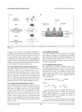
Figure 1. Design of 3D-printed micro-perfused culture (MPC) device. (a) Exploded view of 3D-printed MPC device. (b) Cross-sectional view of 3D- printed
MPC device.
exchange; at the same time, the perfusate collected in a 2.2.6. Scaffold morphology
6-well plate can be removed every other day. The flowrate Electrospun scaffolds were visualized with a Jeol 6340F
used in this study has also demonstrated the benefit of field emission scanning electron microscopy (FE-SEM).
a micro-perfusion setup where medium consumption The samples were imaged at accelerating voltage of 5 kV
could be kept to the minimum without compromising cell and a working distance between 10 and 15 mm. All
support. Therefore, the simulation data should be taken as samples were coated with gold prior to imaging to increase
a reference for maximum allowable flowrate instead of an the sample conductivity.
absolute number to be deployed in this study.
2.2.7. Scaffold porosity and pores
2.2.4. 3D printing of micro-perfused culture device The miniaturized 3D fibrous scaffold porosity was
47
The Asiga Pico2 HD 27 was used in this study for the measured by liquid intrusion method where the weight
fabrication of MPC device. NextDent Ortho Clear, a of dry scaffold was first measured prior to immersion in
biocompatible Class IIa methacrylate UV sensitive resin, 100% ethanol overnight. The scaffold was gently wiped
was used. The 3D-printed MPC device comprises three to remove any excess ethanol on the surface and weighed
distinct layers where each layer has its own functionality again. Scaffold with (n = 3) was calculated with the
(Figure 1). The final optimized processing condition for following equations:
the resin was having a light intensity of 16 mW/cm and an Scaf fold porosity = V eth ×100 %
2
exposure time of 0.45 s with a light wavelength of 385 nm. ( V + V )
pcl
eth
2.2.5. Setup of micro-perfused culture device Where:
The microfluidic perfusion culture was set-up as shown in ( W wet − W ) W dry
dry
Figure 2. A 6-well plate was used to hold the 3D-printed V = ρ eth V = ρ pcl
pcl
eth
MPC device. A syringe pump was used to infuse culture
medium to the MPC device at an optimized rate. The V denotes the volume of ethanol entrapped in the
eth
perfusion culture devices were placed in an incubator scaffold; W and W denote the dry and wet weight
dry
wet
where cells were cultured at 37°C and 5% CO . The of the scaffold, respectively; ρ denotes the density of
eth
2
3
medium outlet which is opposite to the perfusion inlet ethanol (0.789 mg/mm ); ρ denotes the density of
pcl
3
was collected for subsequent cell culture analysis, such as polycaprolactone (1.14 mg/mm ).
Albumin ELISA. The cell inlet port was sealed after the The average pore size of the scaffold was determined by
infusion of cells. measuring at least 50 pores per sample using the ImageJ®
Volume 10 Issue 1 (2024) 146 https://doi.org/10.36922/ijb.0226