
Page 158 - IJB-10-1
P. 158
International Journal of Bioprinting 3D-printed micro-perfused culture device
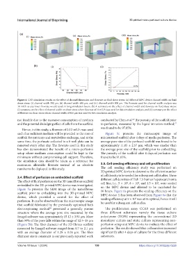
Figure 4. CFD simulation results on the effect of channel dimension and flowrate on fluid shear stress. (a) Effect of MPC device channel width on fluid
shear stress: (i) channel width 300 µm, (ii) channel width 600 µm, and (iii) channel width 800 µm. The flowrate used for channel width analysis was
10 mL/h as any lower flowrate would result in long simulation hours. (b) A summary on the effect of channel width and flowrate on fluid shear stress:
(i) summary on the effect of channel width on shear stress where flowrate of 10 mL/h was used for this simulation analysis, and (ii) summary on the effect
of flowrate on shear stress where channel width of 800 µm was used for this simulation analysis.
not feasible due to the excessive consumption of medium conducted by Chen et al. The porosity of the scaffold prior
45
and the potential dislodging effect of cells from the scaffold. to perfusion, measured by the liquid intrusion method,
47
was found to be 97.85%.
Hence, in this study, a flowrate of 0.12 mL/h was used
such that sufficient medium will be provided to the core of Figure 5c presents the microscopic image of
scaffold for nutrients and metabolites exchange, and at the miniaturized scaffold after 4 days of media perfusion. The
same time, the perfusate collected in a 6-well plate can be average pore size of the perfused scaffold was found to be
removed every other day. The flowrate used in this study approximately 11.05 ± 2.57 µm, which was smaller than
has also demonstrated the benefit of a micro-perfusion the average pore size of the scaffold prior to embedding.
setup where medium consumption could be kept to the The porosity of the scaffold after 4 days of perfusion was
minimum without compromising cell support. Therefore, found to be 91.65%.
the simulation data should be taken as a reference for
maximum allowable flowrate instead of an absolute 3.5. Cell seeding efficiency and cell proliferation
number to be deployed in this study. The cell seeding efficiency study was performed on
3D-printed MPC device to determine the efficient number
of cell density to be seeded for subsequent cell studies. Three
3.4. Effect of perfusion on embedded scaffold different cell densities of Huh 7.5 human hepatocarcinoma
The effect of fluid perfusion on the 3D nanofibrous scaffold cell line, i.e., 5 × 10 , 8 × 10 , and 1.5 × 10 , were seeded
4
6
5
embedded in the 3D-printed MPC device was investigated. on the MPC device and allowed to be incubated for
Figure 5a presents the SEM image of the nanofibrous 16 hours. Figure 6a presents the seeding efficiency on the
scaffold prior to embedding into the 3D-printed MPC MPC device. It has been observed from Figure 6a that the
device, which provided a control for the effect of seeding efficiency at 5 × 10 was at its optimal, hence it will
4
perfusion. It can be observed from the microscopic image be used for subsequent cell studies.
that scaffold fabricated by the previously optimized bath
electrospinning method presented a generally porous The proliferation assay CCK8 was performed on
46
structure where the average pore size measured by the three different substrates namely the tissue culture
ImageJ software was approximately 15.12 ± 3.91 µm. More polystyrene (TCPS) representing the conventional 2D
than 50% of the pore falls within the range of 12.5–15 µm monolayer culture and static culture representing non-
(Figure 5b). The fiber diameter of the 3D fibrous scaffold perfusion setup and MPC device to evaluate the effect of
measured by ImageJ software ranged from 0.7 to 2.1 µm perfusion. The results showed that cell number increased
with an average diameter of 1.28 ± 0.35 µm. The fiber significantly after 4 days of culture for the three different
diameter size is consistent to our previously reported work substrates.
Volume 10 Issue 1 (2024) 150 https://doi.org/10.36922/ijb.0226