
Page 159 - IJB-10-1
P. 159
International Journal of Bioprinting 3D-printed micro-perfused culture device
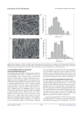
Figure 5. Effect of perfusion on 3D fibrous scaffold. (a) Microscopic image of fibrous scaffold prior to embedding. (b) Percentage pore size distribution of
scaffold prior to embedding. (c) Microscopic image of fibrous scaffold after 4 days of media perfusion on 3D-printed MPC device (without cell seeding).
(d) Percentage pore size distribution of scaffold after 4 days of media perfusion on 3D-printed MPC device (without cell seeding).
3.6. Cell viability studies on 3D-printed there were still dead cells, they were of lesser quantity and
micro-perfused culture device more distributed compared to the static culture. The MPC
To investigate the cell viability of hepatocytes seeded on perfusion culture presented a thicker cell construct with
the 3D-printed MPC device, a LIVE/DEAD assay was more than 400 µm of cell depth observed, demonstrating
performed after 4 days of culture. Figure 7 presents the the benefit of media perfusion on thicker cell constructs.
confocal Z-stack images of the three substrates used in
this study. From the images, Huh7.5 cells cultured on the 3.7. Gene expression and albumin secretion test
TCPS plate presented the least number of dead cells. This The gene expression of albumin and CYP3A cytochrome
is much expected since there is direct medium exchange was performed to examine the function of Huh7.5 cells
on the 2D culture. The static culture, which comprises a cultured on three different substrates. The gene expression
41
miniaturized piece of porous scaffold without perfusion, presented in Figure 8a was normalized to TCPS culture. At
had a relatively good cell infiltration after 4 days of day 4, the expression of albumin was approximately three
culture where cells were found to lie within the scaffold times for MPC platform and relatively marginal expression
of approximately 160-µm thickness from its surface. was detected in the static culture. The albumin gene
However, static culture presents the most number of dead expression of MPC platform was found to be significantly
cells since on a static culture, there bound to be some higher as compared to the other two substrates.
56
regions with limited exchange of nutrients. The condition A significant upregulation of CYP3A7 was observed for
was much improved when perfusion was introduced the MPC platform (~3.4-fold), while the static culture did
to the miniaturized scaffold on MPC device. Although not exhibit any enhancement. The expression of CYP3A7
Volume 10 Issue 1 (2024) 151 https://doi.org/10.36922/ijb.0226