
Page 480 - IJB-10-1
P. 480
International Journal of Bioprinting TPMS bone scaffold
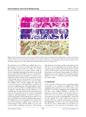
Figure 8. Histological analysis of the bone repair induced by different scaffolds within the mandibular defect model in rabbits. (A) H&E and (B) Masson’s
trichrome staining. (C) ALP immunohistochemical staining. In the images, the alphabet “T” represents a bone scaffold, “arrow” refers to blood vessels,
“yellow star” represents new bone and osteoid, and “green triangle” represents muscles. Notes: IT, TPMS scaffold loaded with I-PRF; NC, negative control;
SIT, TPMS scaffold loaded with I-PRF and SDF-1; ST, TPMS scaffold loaded with SDF-1; T, TPMS scaffold.
the implantation of the TPMS base scaffold, there was a blue represents the nucleus, and the green represents the
greater degree of new bone formation generated around OPG gene. The average fluorescence intensity indicates
the implanted scaffold and also within the scaffold that the expression of OPG is most pronounced in the SIT
structure. Moreover, blood vessels (arrow) could be scaffold, which is higher than other blank control groups
observed distributed throughout the defect site. Scaffolds and positive control groups. Taken together, the collective
groups containing I-PRF or SDF-1 alone or in combination findings in this study indicate the tremendous clinical
substantially increased the formation of new bone tissue utility of SIT and IT scaffolds in promoting mandibular
within the scaffold structure when compared to the base bone defect repair.
scaffold group, with increased quantity of bone lacunae,
osteoblasts, and blood vessel network observed within 4. Conclusion
the scaffolds. These findings suggest the importance
of incorporating these pro-regenerative growth factor In this study, the TPMS bone tissue engineering scaffold
additives in stimulating the uniform formation of dense customized by 3D printing was loaded with I-PRF
bone tissue observed within the scaffold, which is critical containing various growth factors, which was then coated
for effective mandible defect healing. Moreover, in all with SDF-1 to promote cell migration and vascularization.
scaffold groups, no signs of infection or inflammation were The bone tissue scaffold exhibited a structure and
observed, an important factor for effective biomaterial mechanical properties similar to the human cancellous
65
integration and tissue healing. As shown in Figure 8C, bone, and it can recruit cells and continuously release
alkaline phosphatase (ALP) staining showed that all four various growth factors by incorporating SDF-1 with I-PRF
groups of scaffolds were positive, and the positive areas of into the scaffold. The in vitro results showed that the
SIT scaffold were more concentrated in the newly formed developed SIT bone scaffold could promote osteogenesis
bone tissue around the scaffold. Figure 9 indicates the and angiogenesis of cells. In vivo, the findings showed
osteoprotegerin (OPG) staining results of in vivo cells that the SIT bone scaffold induced the formation and
under the effect of different components. DAPI staining vascularization of new bone in the mandibular defect
Volume 10 Issue 1 (2024) 472 https://doi.org/10.36922/ijb.0153