
Page 30 - IJB-2-1
P. 30
Preventing bacterial adhesion on scaffolds for bone tissue engineering
nanoscale consisting of almost vertically aligned na- very similar to the nanostructure of cicada wings as
nocolumns with lengths between 250 and 350 nm with previously reported [13] . Moreover, this kind of dense,
a diameter between 40 and 60 nm, separated (from highly packed nanotopography, together with the separ-
center to center) by 100–200 nm. This nanostructure is ation between nanofeatures can lead to a significant
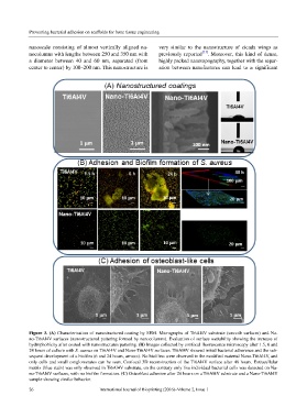
Figure 3. (A) Characterization of nanostructured coating by SEM. Micrographs of Ti6Al4V substrate (smooth surfaces) and Na-
no-Ti6Al4V surfaces (nanostructured pattering formed by nanocolumns). Evaluation of surface wettability showing the increase of
hydrophobicity after coated with nanostructures pattering. (B) Images collected by confocal fluorescence microscopy after 1.5, 6 and
24 hours of culture with S. aureus on Ti6Al4V and Nano-Ti6Al4V surfaces. Ti6Al4V showed initial bacterial adherence and the sub-
sequent development of a biofilm (6 and 24 hours, arrows). No biofilms were observed in the modified material Nano-Ti6Al4V, and
only cells and small conglomerates can be seen. Confocal 3D reconstruction of the Ti6Al4V surface after 48 hours. Extracellular
matrix (blue stain) was only observed in Ti6Al4V substrate, on the contrary only live individual bacterial cells was detected on Na-
no-Ti6Al4V surfaces, with no biofilm formation. (C) Osteoblast adhesion after 24 hours on a Ti6Al4V substrate and a Nano-Ti6Al4V
sample showing similar behavior.
26 International Journal of Bioprinting (2016)–Volume 2, Issue 1